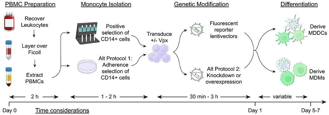
Figure 1. Schematic illustration of monocyte isolation, genetic modification, and differentiation of MDDCs and MDMs.

The major steps of the protocols described in this article are shown (PBMC preparation, CD14+ cell isolation, transduction with lentiviral vectors, and differentiation into MDDCs or MDMs), corresponding with estimates of the time investment required at each step.
