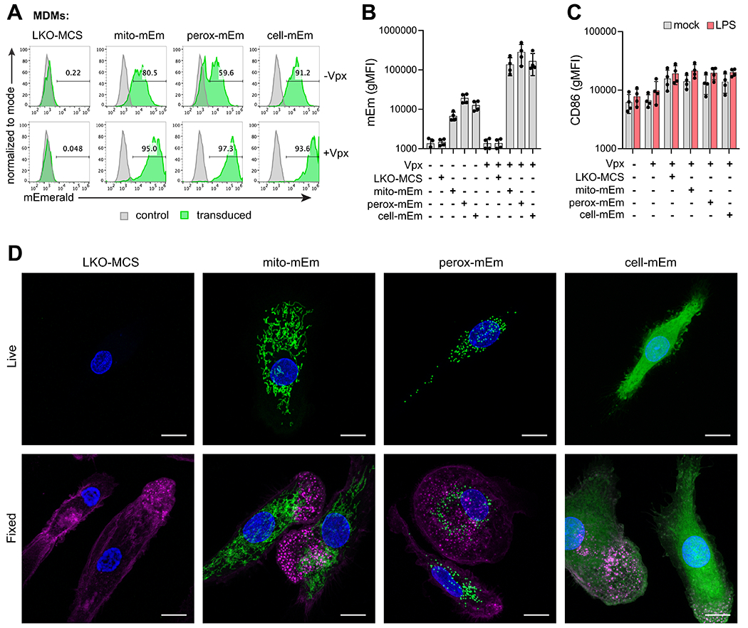
Figure 3. Genetic modification of MDMs with fluorescent lentiviral reporter vectors.

A) Representative flow cytometry histograms of mEmerald expression in MDMs at day 7 after transduction with LKO-MCS (vector alone), mito-mEm, perox-mEm, or cell-mEm in the presence or absence of SIV-VLPs packaging Vpx. B) Geometric mean fluorescence intensity (gMFI) of mEmerald expression in transduced MDMs. C) gMFI of CD86 expression in mock-treated or transduced MDMs that were either unstimulated or stimulated with LPS (100 ng/ml for 24 h). For (C) and (D), n = 4 independent donors. D) Representative z-stack projections (merge of 10-20 planes, 0.5 micron step size) of live and fixed images of transduced MDMs acquired with an LSM880 Airyscan Fast confocal microscope. Images reveal localization of fluorescent markers (mEmerald expression, green), nuclei (DAPI, blue), and actin (Phalloidin, magenta – only depicted in fixed cell images). Scale bar = 10 μm.
