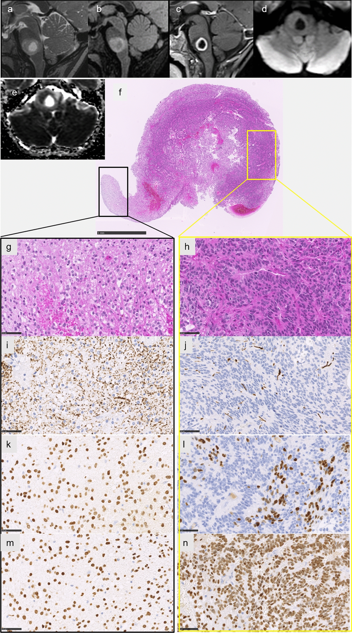
Fig. 4.

Radiological and histopathological features of case #6. Sagittal T2-weighted image (a) and FLAIR image (b) showing an infiltrative lesion of the pons with a central liquid part consistent with necrosis, with a peripheral enhancement of the necrosis on T1-weighted image with gadolinium injection (c), and intermediate signal on diffusion-weighted image (d) with partially restricted diffusion on apparent diffusion coefficient map (e). The biopsy highlighted two different histopathological components (f HPS, magnification × 50). The glial diffuse component (g HPS, magnification 400), associated with an ependymal component with rosettes (h HPS, magnification 400). Neurofilament immunostaining confirming the infiltrative pattern in the glial component (i magnification 400) and a more circumscribed pattern in the ependymal component (j magnification 400). Diffuse immunopositivity for Olig2 in the glial component (k magnification × 400), and partial expression of Olig2 in the ependymal component (l magnification × 400). Diffuse immunoexpression of H3K27M in both tumoral components (m–n magnification 400). Scale bars represent 1 mm (f) and 50 µm (g–n). HPS: Hematoxylin Phloxin Saffron
