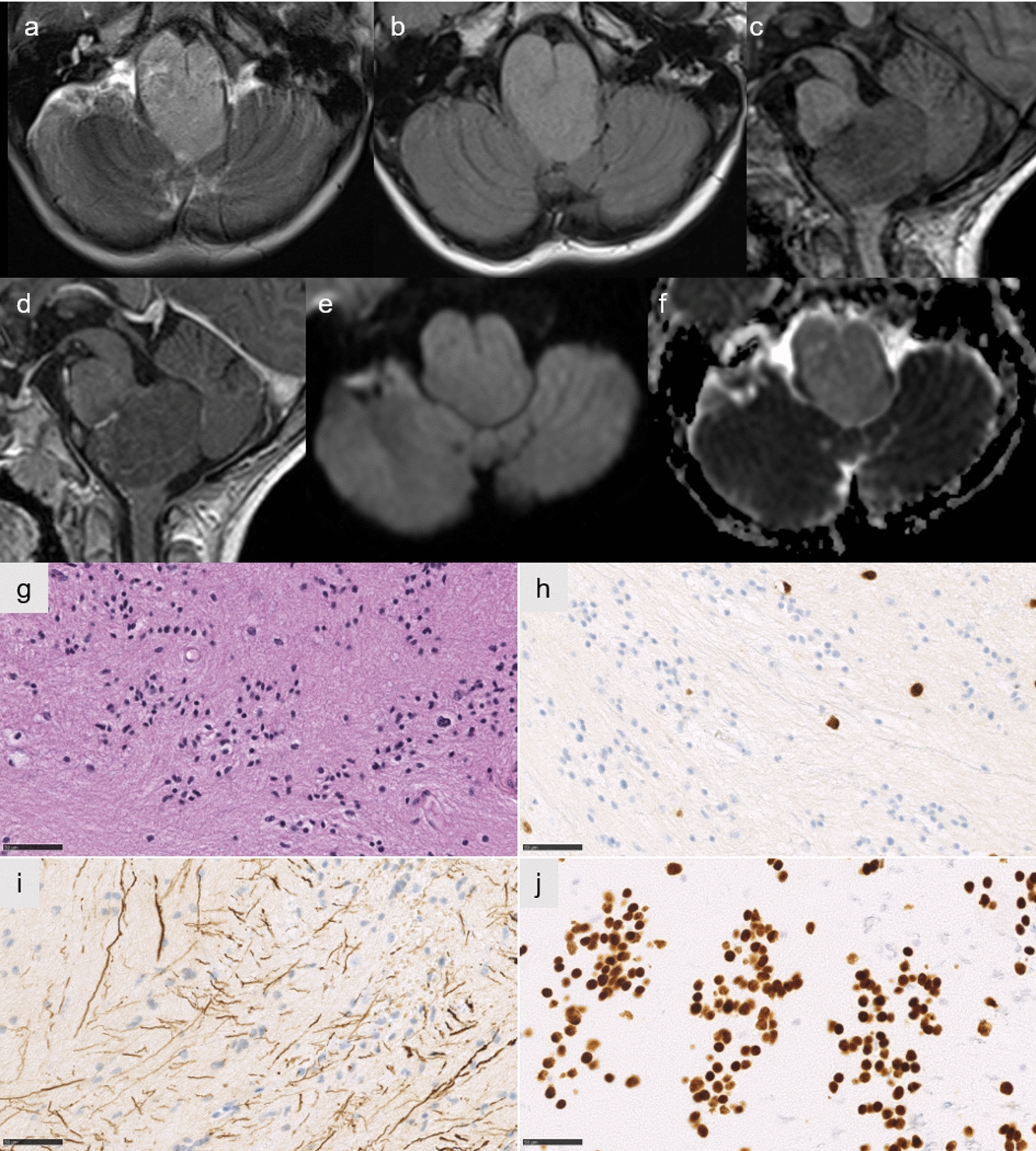
Fig. 5.

Radiological and histopathological features of case #9. Axial T2-weighted and FLAIR image showing a circumscribed lesion of the medulla oblongata (a,b), without enhancement on sagittal T1-weighted image with gadolinium injection (c before injection, d after injection), and low signal on diffusion weighted image (e) with increased diffusion on apparent diffusion coefficient map (f). A paucicellular tumor with subependymal features (g HPS, magnification 400). No immunoreactivity for Olig2 except in residual glial cells (h HPS, magnification 400). Neurofilament immunostaining showing an infiltrative pattern (i magnification 400). Diffuse immunoexpression of H3K27M in tumor cells (j magnification 400). Scale bars represent 50 µm. HPS: Hematoxylin Phloxin Saffron
