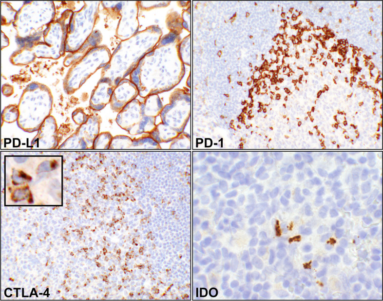
Fig. 1.

Controls for the immunohistochemical (IHC) reactions. (PD-L1) A photomicrograph showing intense predominant staining of syncytiotrophoblasts appearing as a linear reaction. Tonsils can be also used as control for this immune checkpoint (20 × objective). (PD-1) A portion a lymphoid follicle in tonsil with adjacent germinal center depicting T-helper cells by IHC (20 × objective). The reaction is on cell membrane of the cells. (CTLA-4), Photomicrograph of a segment of a tonsillar germinal center with the adjacent follicular cell (20 × objective). IHC has detected the activated T-cells where the reactions are coarsely granular and intracytoplasmic (inset, 60 × objective, digitally magnified). (IDO) A focus of tonsillar lymphoid tissue showing a few dendritic cells with granular intracytoplasmic reaction (60 × objective). Abbreviations: PD-L1, programmed death-ligand 1; PD-1, programmed cell death protein 1; CTLA-4, cytotoxic T lymphocyte antigen 4; IDO, indolaimine-2, 3-deoxygenase
