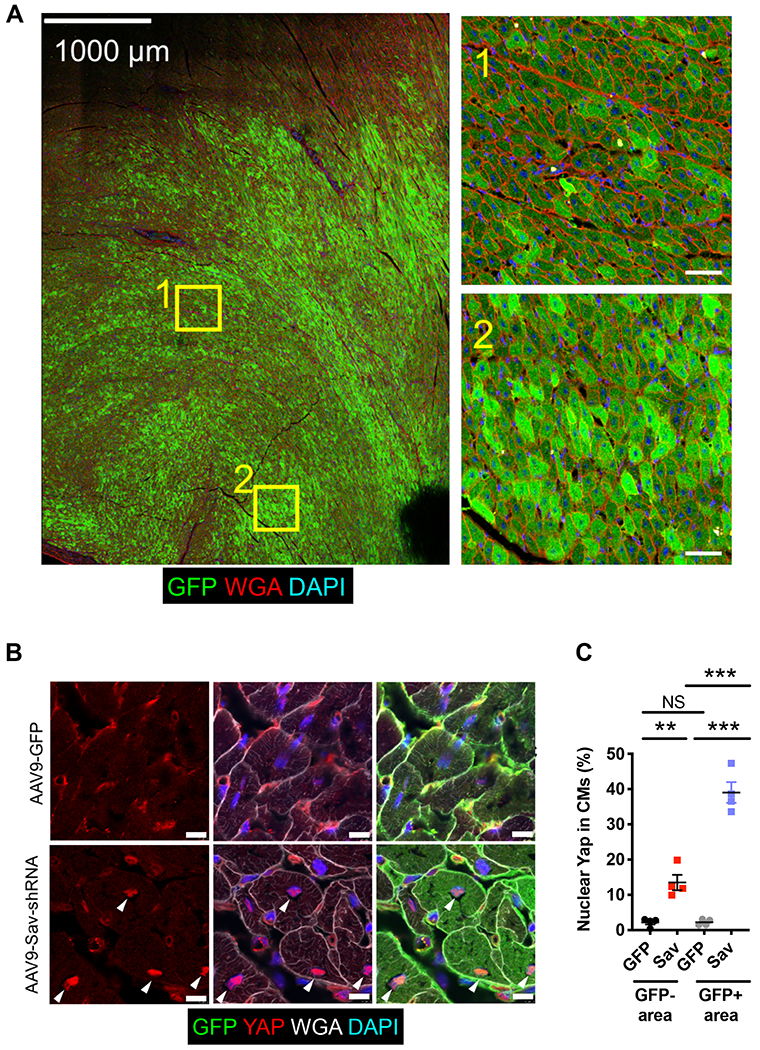
Fig. 1. Increased Yap nuclear localization in cardiomyocytes after AAV9-Sav-shRNA gene therapy.

(A) Shown is a representative tiled image of GFP staining of a section from an AAV9-Sav-shRNA–injected pig heart (pig P-1902; euthanized at 104 days and myocardial infarction, 33 days after viral vector injection). Inset images on the right show the magnification of the boxed areas in the tile image on the left. Scale bars, 1000 μm (left image) and 50 μm (right inset images). DAPI, 4′,6-diamidino-2-phenylindole. (B) Immunofluorescence staining for endogenous GFP and Yap shows the subcellular localization of Yap in GFP-positive cardiomyocytes that received AAV9-Sav-shRNA gene therapy. White arrowheads indicate nuclear Yap. Scale bars, 10 μm. (C) Quantification of the percentage of nuclear Yap in GFP-positive and GFP-negative cardiomyocytes of pig hearts injected with AAV9-Sav-shRNA (Sav) or AAV9-GFP (GFP) as control (n = 4 per group). Data were compared using one-way ANOVA with Tukey’s post hoc test. Data are presented as the mean ± SEM. **P < 0.01 and ***P < 0.001; NS, not significant.
