
Fig. 1.
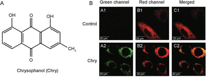
Detection of Chry in mitochondria of HepG2 cells by laser confocal microscopy. (A) The chemical structure of Chry. (B) Fluorescent imaging of mitochondria. The cells were incubated with 50 μmol/L of Chry for 15 min, and then stained with MTDR (an exclusive dye for mitochondria), for another 15 min.