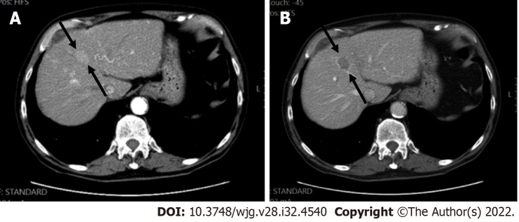
Figure 5.
Hepatocellular carcinoma in a 60-year-old man with hepatitis C. A: Computed tomography scan, late arterial phase were the hepatic artery and portal vein are both enhanced, showed a segment VIII lesion measuring 27 mm with non-rim arterial phase hyper-enhancement; B: In the portal phase, washout was seen.

