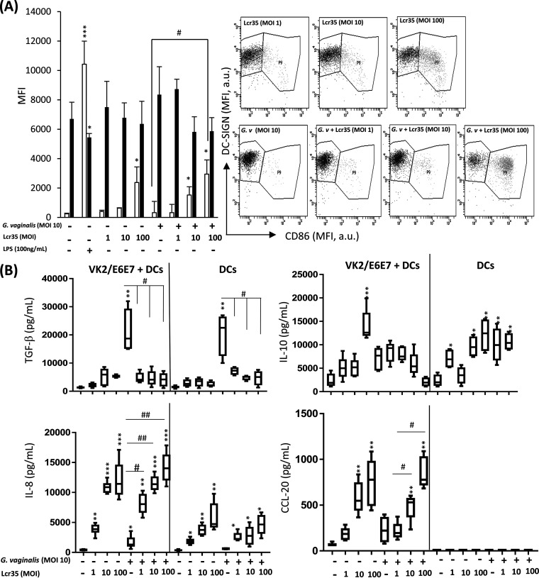
FIG 1.
Effect of Lcr35 on G. vaginalis-infected dendritic cells in a coculture model. Human monocyte-derived dendritic cells were exposed indirectly through the vaginal epithelial cell monolayer to UV-inactivated G. vaginalis (MOI, 10) alone or UV-inactivated Lcr35 (MOI, 1 to 100) for 48 h. (A) The effects of DC functional maturation were determined by measuring the DC-surface expression of DC-SIGN (black bars) and CD86 (white bars) by flow cytometry. The dot plots and histograms show MFI values on gated DCs. DC-SIGN/CD86 dot plots gated on human DCs. (B) The secretions of cytokines TGF-β, IL-10, IL-8, and CCL-20 were measured in the supernatant of coculture VK2/E6E7 + DC (lower part) or DC alone by ELISA. Values are the means ± SEM; n = 4 to 5; *, P < 0.05; **, P < 0.01 compared with noninfected DCs. #, P < 0.05; #, P < 0.01 compared with DCs infected with G. vaginalis.

