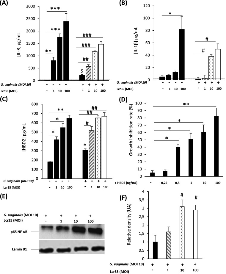
FIG 2.
Effect of Lcr35 on innate immune response in G. vaginalis-infected vaginal epithelial cells. VK2/E6E7 cells were infected with G. vaginalis (MOI, 10) alone or with Lcr35 at different MOIs (MOI, 1 to 100). IL-8 (A), IL-1β (B), and HBD-2 (C) concentrations were analyzed 6 h postinfection by ELISA. The detection limits of IL-8 and IL-1β were 312 and 39 pg/mL, respectively. (D) Growth inhibition rates of G. vaginalis after treatment with various concentrations of HBD-2 for 4 h. (E) The presence of the p65 NF-κB and Lamin B1 proteins were detected by Western blotting in cellular extracts of VK2/E6E7 cells stimulated for 3 h with G. vaginalis alone or with Lcr35. (F) Densitometric analysis of the data in Fig. 2E by using Image Lab 2.0 software (n = 3). Representative data of 3 to 8 independent experiments. Values are the means ± SEM; *, P < 0.05; **, P < 0.01 compared with uninfected cells. #, P < 0.05; ##, P < 0.01 compared with G. vaginalis-infected cells.

