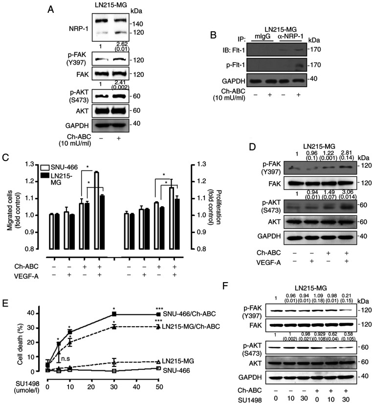
Figure 5.
Effect of eliminating chondroitin sulfate modification on NRP-1 in GB cells. (A) Elimination of chondroitin sulfate modification on NRP-1 using Ch-ABC (10 mU/ml) treatment in LN215-MG cells, after which the cell lysates were subjected to western blot analysis using antibodies specific for NRP-1, p-FAK (Y397), total FAK, p-AKT (S473), and total AKT. GAPDH was used as a control. The relative pixel intensities of the target molecules were assessed by densitometric analysis using ImageJ analysis software. Data are representative of three individual experiments. (B) An increase in interaction between NRP-1 and Flt-1 was determined by immunoprecipitation using 1% NP-40 lysis buffer in LN215-MG cells following Ch-ABC treatment. The relative pixel intensities of the target molecules were assessed by densitometric analysis using ImageJ analysis software. GAPDH was used as a control. Data are representative of three individual experiments. (C) SNU-466 and LN215-MG cells incubated with or without Ch-ABC in the absence or presence of VEGF-A (50 µg/l) for 4 h (migration) or 72 h (proliferation). VEGF-mediated cell migration was assessed using Transwell migration assay (left) and proliferation was evaluated by WST-1 assay (right) (P-values were evaluated with Student's t-tests). (D) LN215-MG pretreated with or without Ch-ABC for 24 h in the absence or presence of VEGF-A (50 µg/l) for 120 min, after which the LN215-MG cell lysates were subjected to western blot analysis using antibodies specific for p-FAK (Y397), total FAK, p-AKT (S473), and total AKT. GAPDH was used as a control. The relative pixel intensities of the target molecules were assessed by densitometric analysis using ImageJ analysis software. Data are representative of three individual experiments. (E) SNU-466 and LN215-MG cells were incubated with SU1498 in a dose-dependent manner in the absence or presence of Ch-ABC for 24 h. Cell death was assessed using an LDH assay (n=3; Tukey's post hoc test was applied to significant group effects in ANOVA; *P<0.05 and ***P<0.005). (F) LN215-MG cells were incubated with SU1498 in a dose-dependent manner in the absence or presence of Ch-ABC for 24 h, after which the cell lysates were subjected to western blot analysis using antibodies specific for p-FAK (Y397), total FAK, p-AKT (S473), and total AKT. GAPDH was used as a control. The relative pixel intensities of the target molecules were assessed by densitometric analysis using ImageJ analysis software. Data are representative of three individual experiments. NRP-1, neuropilin-1; GB, glioblastoma; Ch-ABC, chondrotinase ABC; p-, phosphorylated; Flt-1, FMS related receptor tyrosine kinase 1; GAPDH, glyceraldehyde 3-phosphate dehydrogenase; VEGF-A, vascular endothelial growth factor-A.

