
Figure 4. dMLH-mediated DNA sensing promotes T-cell priming independent of TMB.
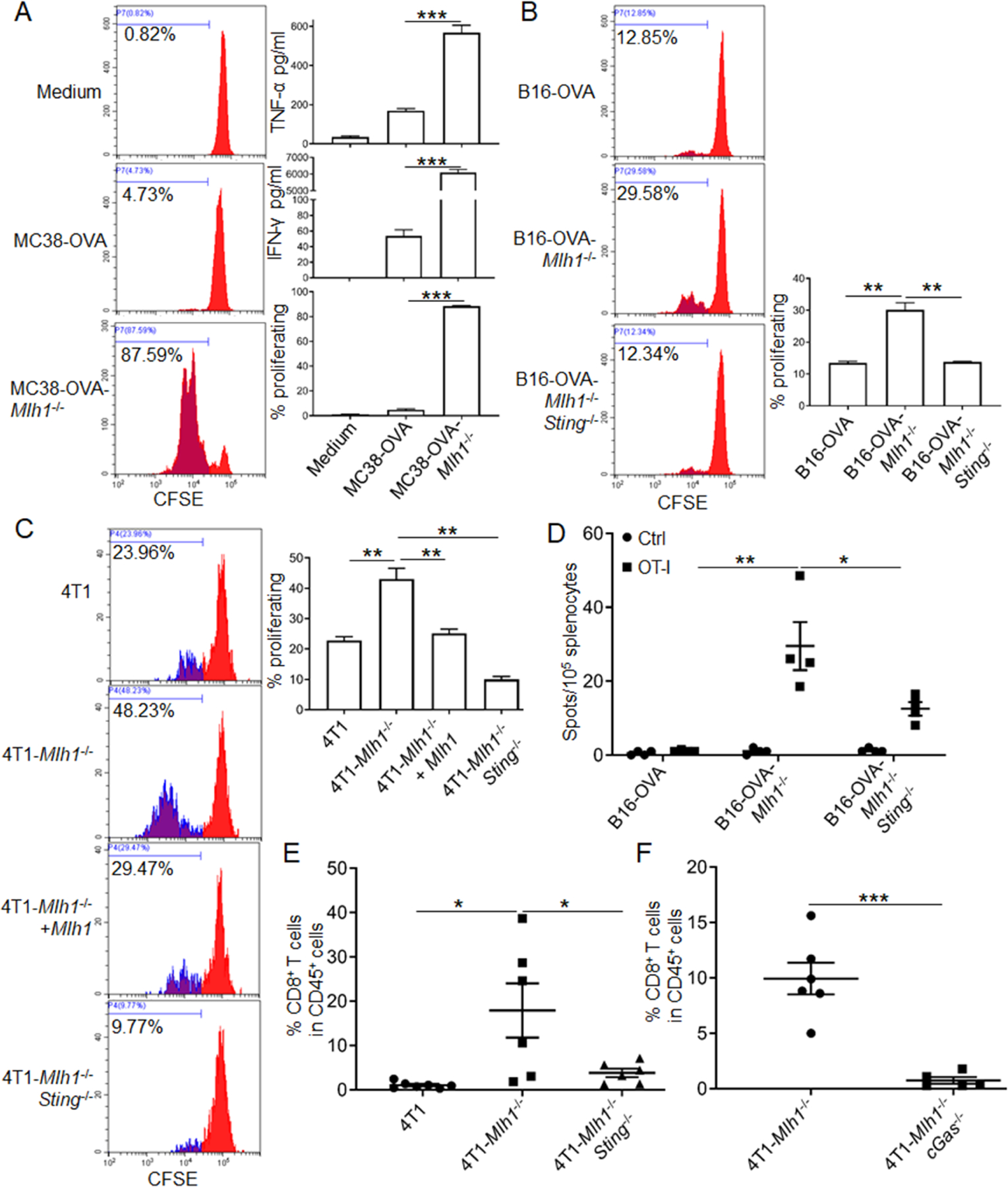
(A and B) BMDCs pre-educated with MC38-OVA and B16-OVA cells were co-cultured with OT-I T cells, then T-cell proliferation was determined. IFN-γ and TNF-α were quantified. Representative FACS histograms and statistic data are shown (also see Figures S4A and S4B).
(C) Supernatants were added into co-culture system of BMDCs and OT-I T cells, then T-cell proliferation was determined. Representative FACS histograms and statistical data are shown.
(D) B16-OVA cells were inoculated into WT C57BL/6 mice (n=4). One week later, cells from the spleen were isolated and re-stimulated by OT-I peptide or control SIY peptide in vitro. T-cell responses were determined by IFN-γ ELISPOT assay.
(E and F) Fragmented tumor tissues derived from 4T1 cells with d Mlh1 (D129), double deficiency cells of Mlh1 plus Sting (D136), and double deficiency cells of Mlh1 plus cGAS (D144) were implanted into WT BALB/C mice (n=6–7). Eleven days later, tumor-infiltrating T cells were detected by FACS.
Data are represented as mean ± SEM. Unpaired t test was used to determine significance.