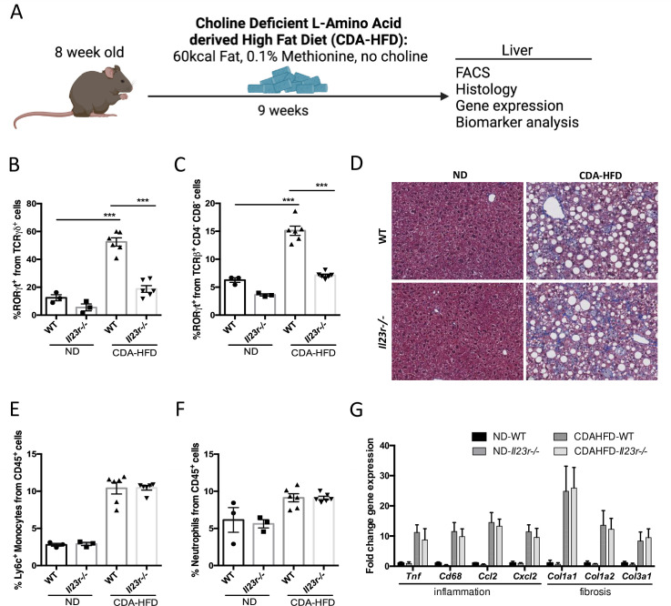
Fig 6. IL-23 signaling does not contribute to liver inflammation and fibrosis in the CDA-HFD NASH model.
WT and Il23r-/- mice were fed CDA-HFD for 9 weeks, followed by liver analysis (A). Percent quantification of hepatic RORγt in γδ T Cells (B) and MAITs (C). Trichrome staining images and quantification of Inflammation (E) and Trichrome Collagen content (F). Liver mRNA expression of Tnf, Cd68, Ccl2, Cxcl2, Col1a1, Col1a2, and Col3a1 (G). Groups: ND WT n = 3, ND Il23r-/- n = 3, CDA-HFD WT n = 6, CDA-HFD Il23r-/- n = 6. Data represents mean ± S.D. ***p < 0.0005, one-way ANOVA.

