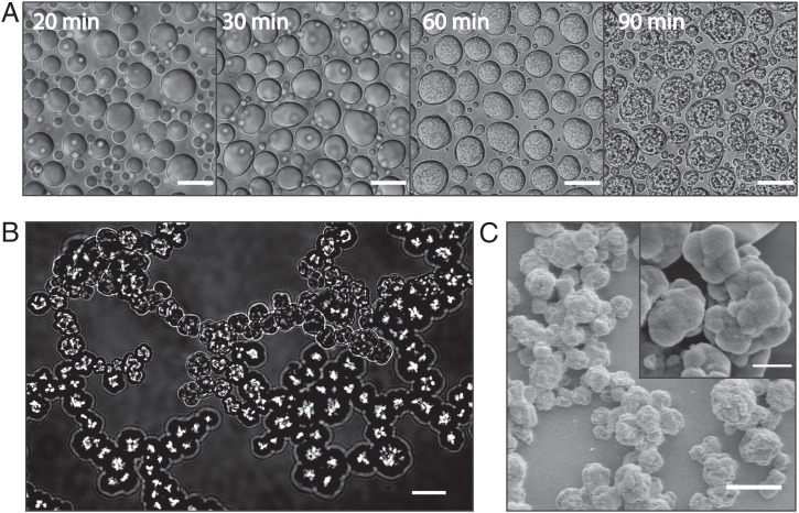
Fig. 2.
Minielastin maturation from liquid coacervates into solid form. (A) Maturation of minielastin condensates over the course of 90 min at 23 °C. (Scale bars: 10 μm.) (B) A matured minielastin sample formed in the capillary flow chamber (Movies S1 and S2). (Scale bar: 20 μm.) (C) Scanning electron microscopy images of mature minielastin obtained after overnight incubation at 37 °C. (Inset) Enlarged image of mature minielastin droplets. Protein concentrations in DIC and scanning electron microscopy images are 130 and 230 μM, respectively. (Scale bars: C, 7 μm; C, Inset, 2 μm.)

