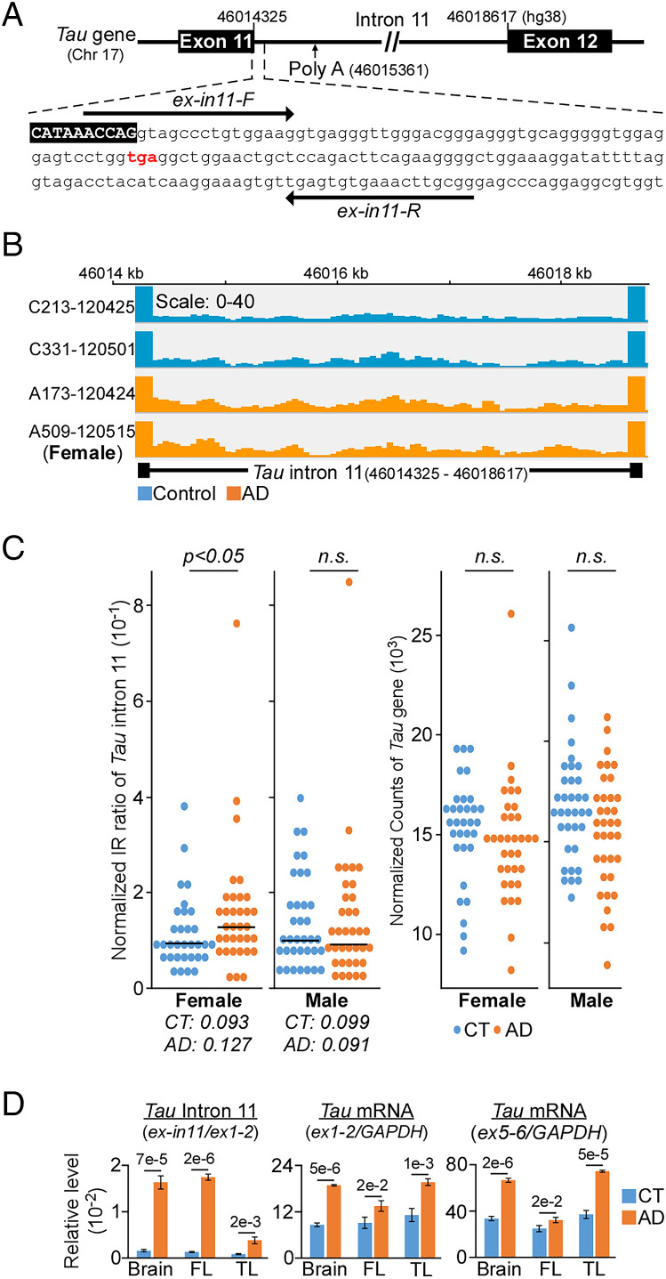
Fig. 1.
Increased retention of intron 11 of Tau gene in AD female. (A) Intron 11 contains a premature stop codon (red) with a canonical polyadenylation site that is located 1 kb downstream. Arrows indicate qPCR primers. (B) Integrative genomic viewer of intron 11 from control (blue) and AD (orange) patients. (C, Left) Statistically significant higher intron retention (IR) is observed only in female AD dorsal lateral prefrontal cortex (n = 34) as compared to control (CT, n = 32). Each dot represents individual normalized IR ratio. Right: No differential expression in the Tau gene between CT and AD cohorts. (D) qPCR validation of intron 11 (relative to Tau exon1/exon2) and Tau expression (exon1/exon 2 and exon5/exon6 relative to GAPDH) from three distinct pairs of brain tissues. FL, frontal lobe; TL, temporal lobe. Data presented as mean of triplicates ± SD, P values calculated by two-tailed t test.

