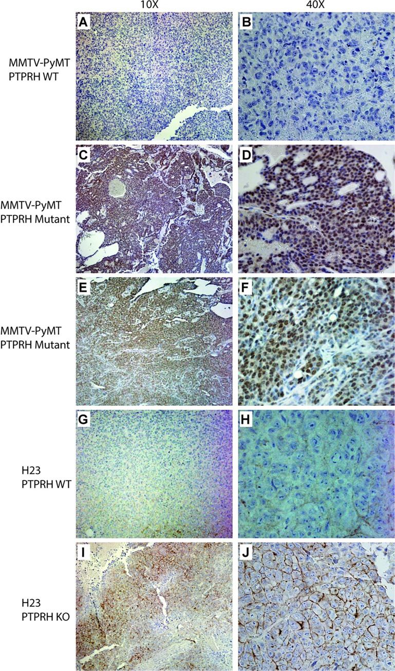
Fig 5. Phospho-EGFR Immunohistochemistry reveals nuclear and membrane staining.
A MMTV-PyMT tumor that was wild type for Ptprh was used in immunohistochemistry for phosphoEGFR revealing essentially no staining at low (10x) and high (40x) magnification (A and B respectively). A PyMT tumor with a V483M PTPRH mutation revealed largely nuclear staining across the entire tumor and was reflective of these tumors. This was repeated in a second tumor with identical results (E and F). IHC for pEGFR in the H23 parental line with wild type PTPRH revealed low levels of expression, largely in the membrane (G and H). The H23 PTPRH CRISPR knockout human tumor line grown in mice revealed membrane specific staining for phosphoEGFR (I and J).

