. 2021 Oct 26;10:e66039. doi: 10.7554/eLife.66039

Figure 1. The central complex (CX) and accessory brain regions.

(A) The portion of the central brain (aquamarine) that was imaged and reconstructed to generate the hemibrain volume (Scheffer et al., 2020) is superimposed on a frontal view of a grayscale representation of the entire Drosophila melanogaster brain (JRC 2018 unisex template [Bogovic et al., 2020]). The CX is shown in dark blue. The midline is indicated by the dotted black line. The brain areas LO, ME, and SEZ, which lie largely outside the hemibrain, are labeled. (B) A zoomed-in view of the hemibrain volume, highlighting the CX and accessory brain regions. (C) A zoomed-in view of the structures that make up the CX, the ellipsoid body (EB), protocerebral bridge (PB), fan-shaped body (FB), asymmetrical body (AB), and paired noduli (NO). (D) The same structures viewed from the lateral side of the brain. (E) The same structures viewed from the dorsal side of the brain. The table below shows the abbreviations and full names for most of the brain regions discussedin this paper. See (Scheffer, 2020) for details. Anatomical axis labels: d: dorsal; v: ventral; l: lateral; m: medial; p: posterior; a: anterior.

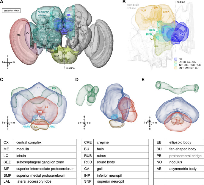
Figure 1.

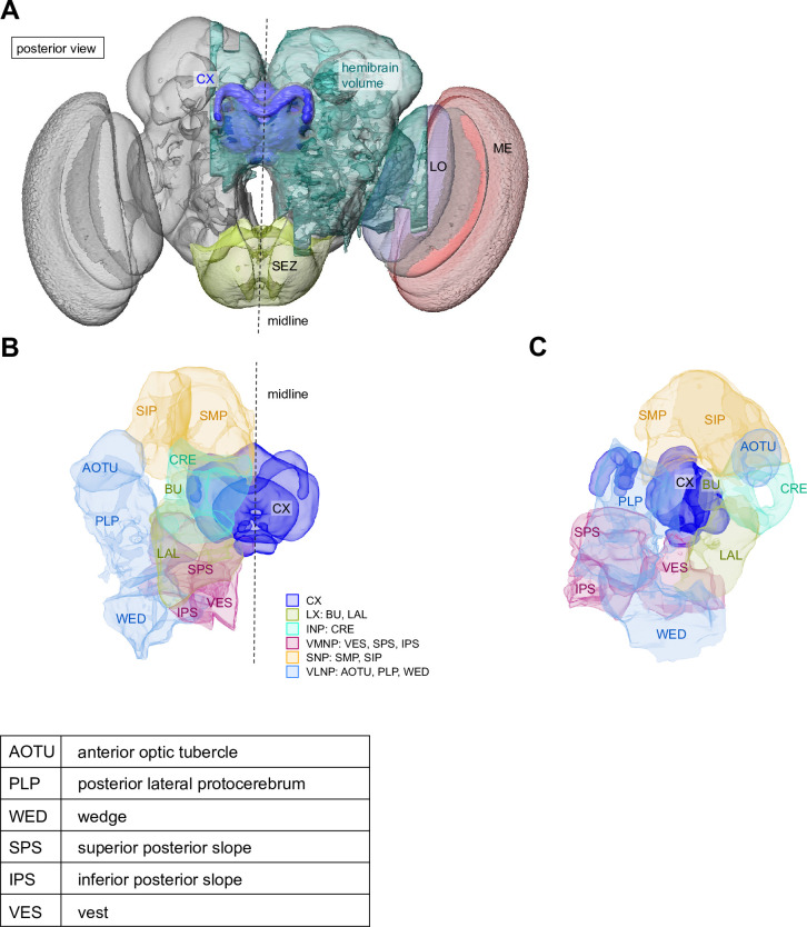
Figure 1—figure supplement 1. The central complex (CX) and additional accessory brain regions.

Figure 1—figure supplement 1.

(A) Posterior view of the hemibrain volume shown in Figure 1A. (B) A zoomed-in view of the CX and additional accessory brain regions not shown in Figure 1. (C) Lateral view of (B).

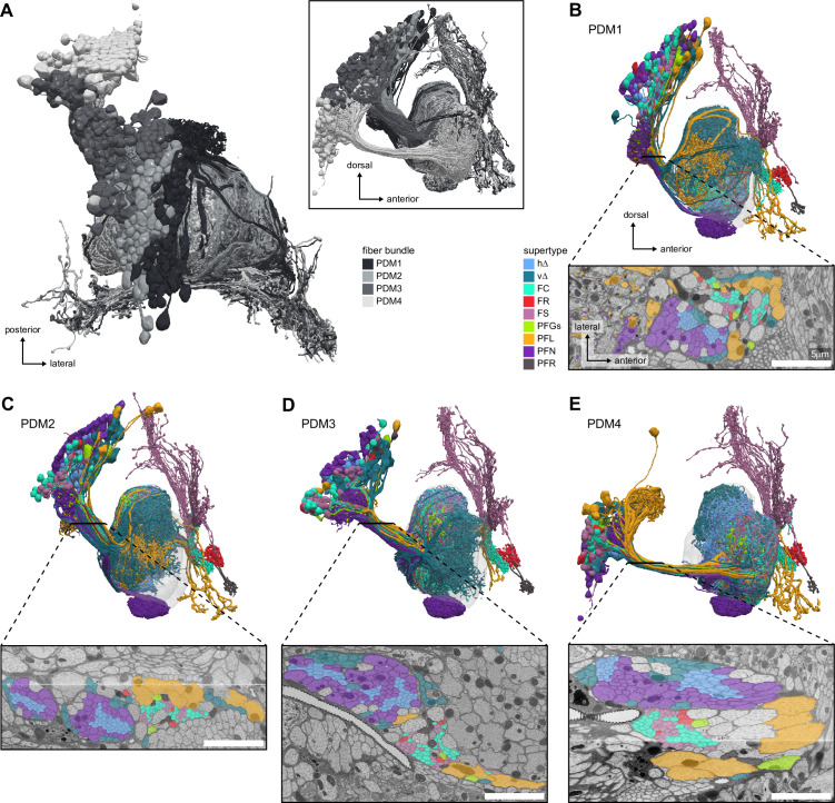
Figure 1—figure supplement 2. Fan-shaped body (FB) neurons tracts.

Figure 1—figure supplement 2.

(A) Top and side (inset) views of the PDM1 to PDM4 cell clusters, corresponding to the DM1 to DM4 hemilineages, also known as w,x,y,z hemilineages in other insect species (Boyan and Williams, 2011; Izergina et al., 2009; Williams, 1975). These cell clusters encompass all FB types except the tangential FB neurons. (B–E) Lateral view of the FB neurons in the PDM clusters (top) and an EM cross-section of the bundle of processes connecting their somata to the FB (bottom). h∆ and v∆ neurons travel with the PFN neurons, but have neurites with much smaller diameters. FC, FS, and FR neurons travel in between the PFN and PFL neurons, and generally have small-diameter processes. Scale bar 5 μm.

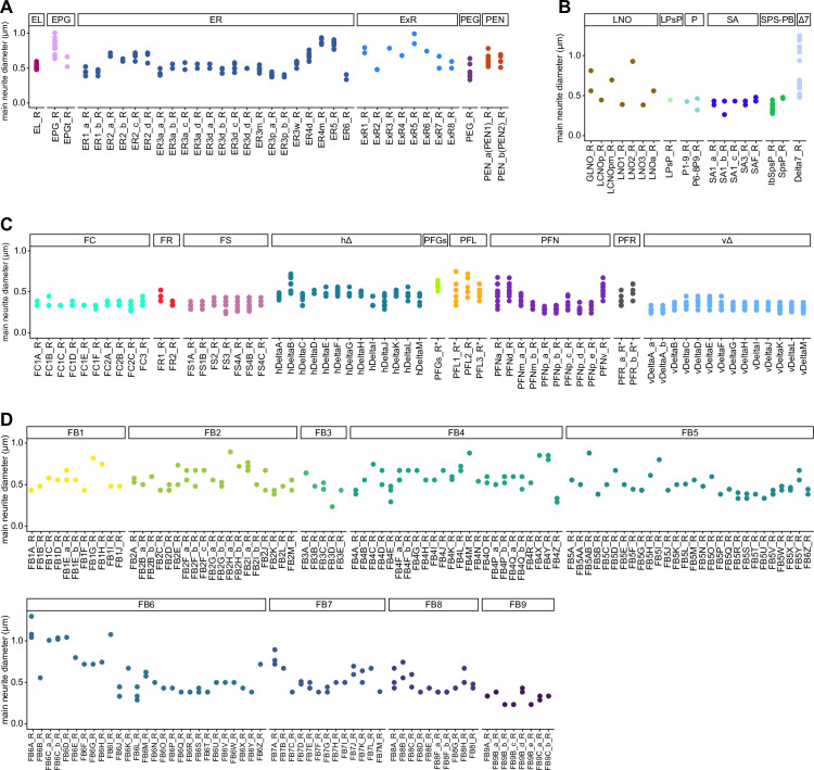
Figure 1—figure supplement 3. Main neurite diameter of central complex (CX) neurons.

Figure 1—figure supplement 3.

Median diameter of the processes between the somata and main branchpoints of all CX neurons, grouped by type. Each point is a neuron, each x-coordinate a type. Note that there is some variability in the detection of the main branchpoint of neurons. (A) EB neurons (B) PB, NO and SA neurons (C) FB neurons (except FB tangentials) (D) FB tangential neurons.