. 2021 Oct 26;10:e66039. doi: 10.7554/eLife.66039

Figure 14. Overview of extrinsic ring (ExR) neurons.

(A) Region arborization plot of all ExR types from the right hemisphere, showing average pre- and postsynaptic counts by region. Indicated below the plot is a qualitative categorization into three groups: mostly input to the ellipsoid body (EB) (blue), mostly output from the EB (pink), and mixed (black). (B) Similarity matrices (see Materials and methods) for ExR neurons based on all their inputs (Bi) and outputs (Bii). ExR-type labels are colored according to groups in (A). (C) Type-to-type connectivity matrix of ExR to EB columnar neurons (Ci) and EB columnar to ExR neurons (Cii).

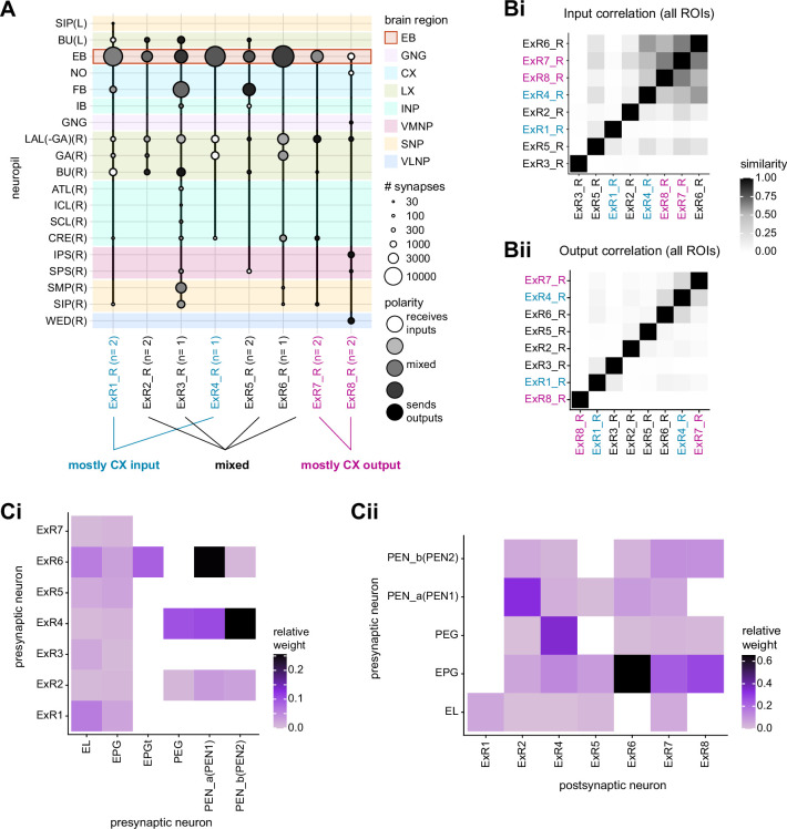
Figure 14.

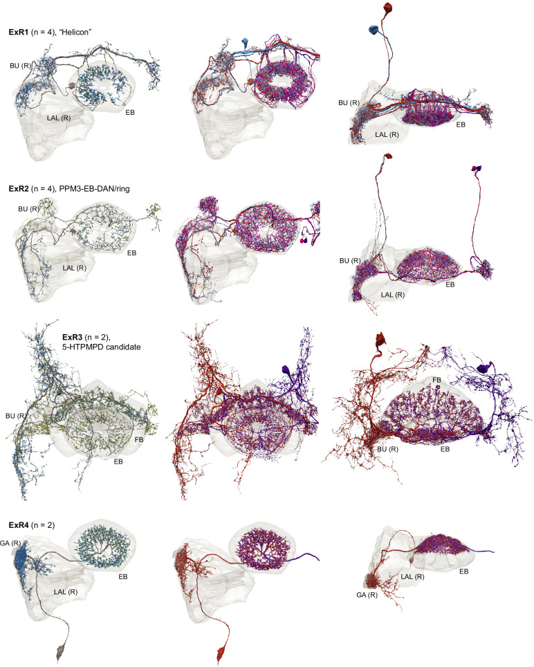
Figure 14—figure supplement 1. Morphological renderings of all ExR types: ExR1, ExR2, ExR3, and ExR4.

Figure 14—figure supplement 1.

Some of the innervated neuropils are shown. The left column shows a single, right hemisphere ExR neuron for each type with presynaptic sites marked by yellow dots and postsynaptic sites marked by blue dots. The middle and right columns show morphological renderings of the complete population.
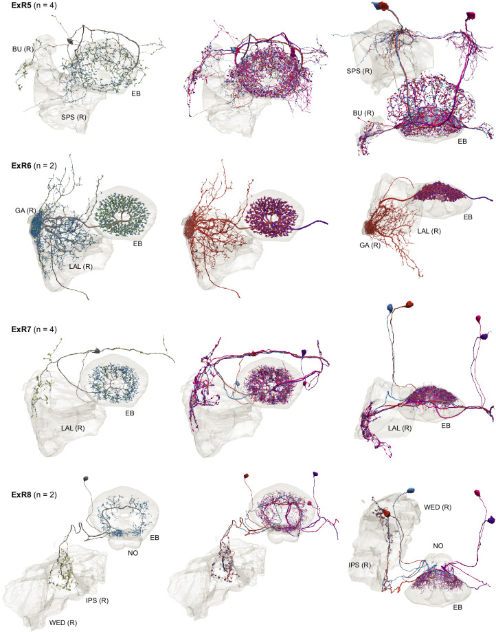
Figure 14—figure supplement 2. Morphological renderings of ExR type. ExR4, ExR5, ExR6, ExR7, ExR8.

Figure 14—figure supplement 2.

See Figure 14—figure supplement 1 for details on presentation.
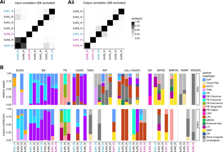
Figure 14—figure supplement 3. Comparison of inputs and outputs of extrinsic ring (ExR) neurons.

Figure 14—figure supplement 3.

(A) Similarity matrices (see Materials and methods) as in Figure 14B but excluding inputs in the ellipsoid body (EB) (Ai) or outputs in the EB (Aii). (B) Stacked bar graph illustrating the fraction of inputs from and outputs to extrinsic ring (ExR) partners, grouped into supertypes and separated by brain region. Inputs and outputs are normalized per neuron type and brain region. The connectivity strength for inputs and outputs is measured by relative weight and output contribution, respectively. ExR-type labels are colored according to groups in Figure 14A.
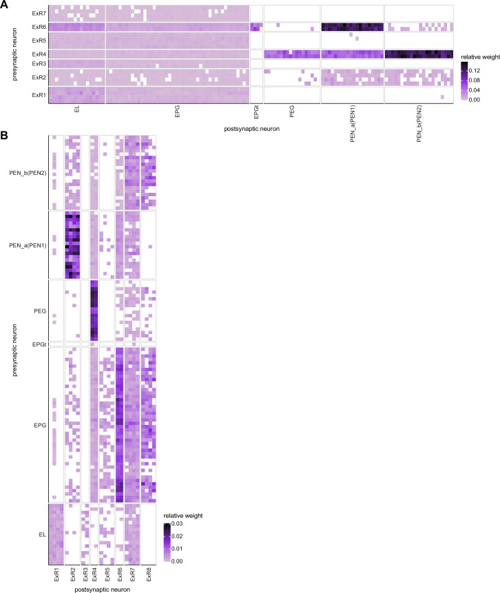
Figure 14—figure supplement 4. Neuron-to-neuron connectivity matrices for connections between extrinsic ring (ExR) and columnar neurons in the ellipsoid body (EB).

Figure 14—figure supplement 4.

(A) Connections from ExR to columnar EB neurons. (B) Connections from columnar EB neurons to ExR.