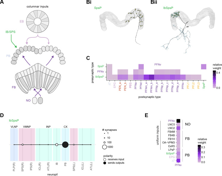
Figure 22. Protocerebral bridge (PB) input and inner neuron connectivity to output neurons.
(A) Schematic depicting the neuropil that bring input to the PB via columnar neurons that target single PB glomeruli. (B) Morphological renderings of single SpsP (Bi) and IbSpsP (Bii) neurons. (C) Type-to-type connectivity matrix from select PB inputs (IbSpsP, PFNv, and SpsP neurons) to PB output neurons. The SpsP neurons also connect to themselves. (D) Region arborization plot for the right IbSpsP neurons. The left IbSpsP neurons were not fully contained in the imaged volume. (E) Type-to-type inputs to the PFNv neurons, separated by neuropil region.
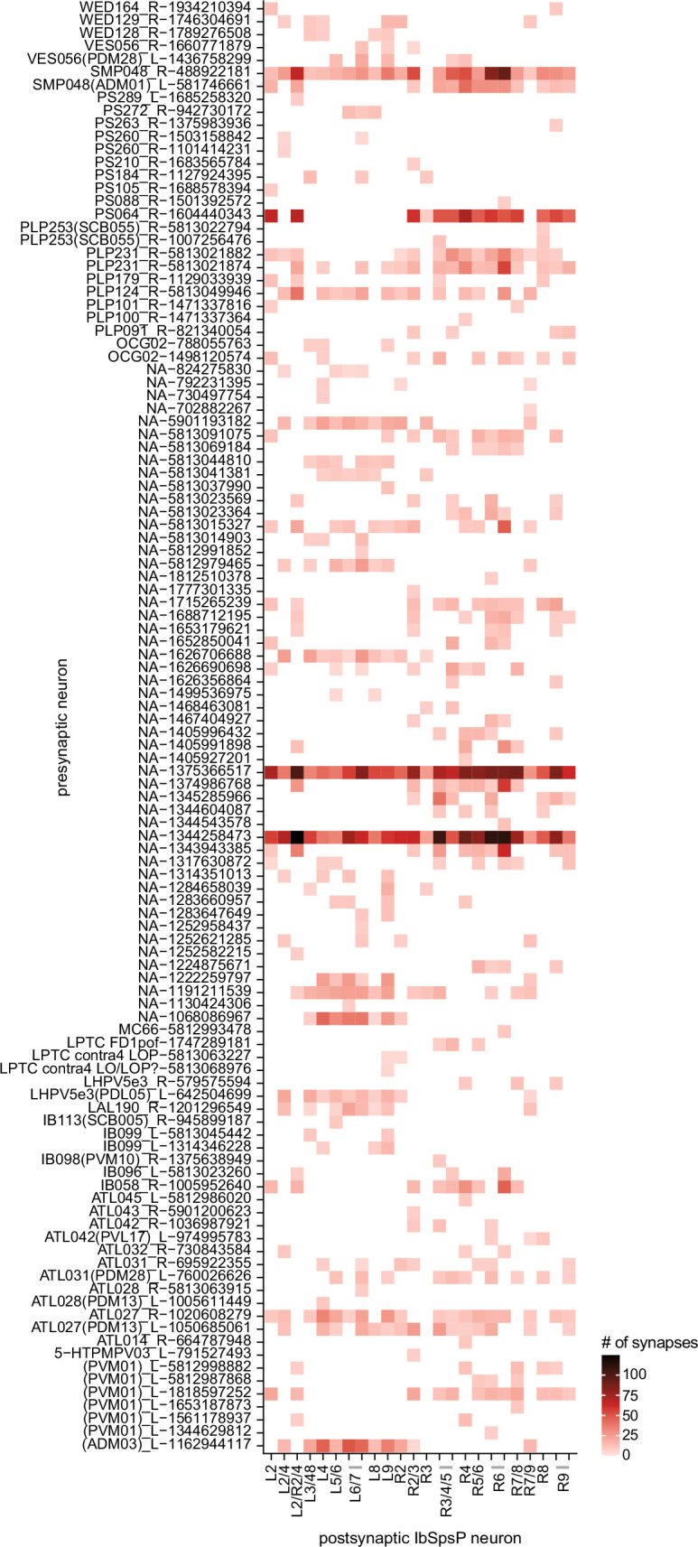
Figure 22—figure supplement 1. Presynaptic partners of the IbSpsP neurons, outside of the protocerebral bridge (PB).


