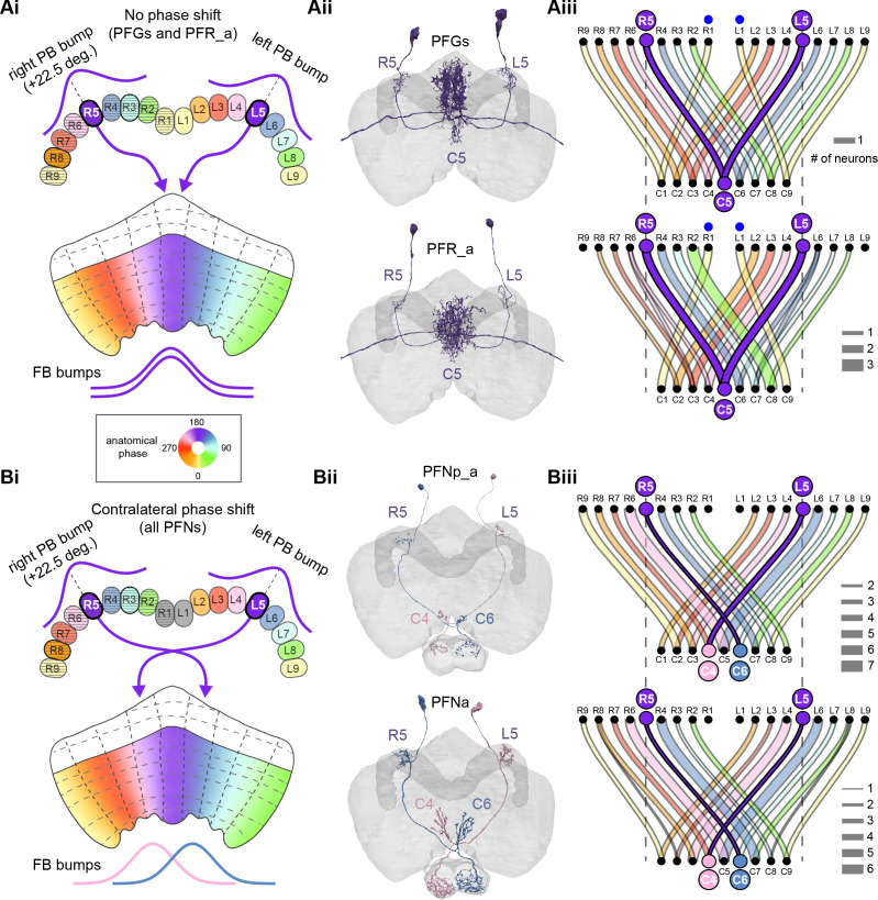
Figure 30. PB-FB-* neurons have type-specific phase shifts in fan-shaped body-to-protocerebral bridge (PB-to-FB) projections.
(A) PFGs and PFRa neurons connect PB glomeruli to FB columns with no phase shift. (Ai) Schematic of a PB-to-FB projection pattern with no phase shift. PB glomeruli and FB columns are colored according to anatomical phase. Based on EB-to-PB columnar neuron projection patterns (EPG neurons, see Figure 16), when a bump is centered at L5 in the left PB, a second bump will be centered between R5/R4 in the right PB (both marked in purple). With no phase shift in their projection pattern, neurons innervating R5/L5 both project to C5 in the FB. This pattern, repeated across glomeruli/columns (see Aiii), would bring the two bumps in the PB to approximately the same FB location. (Aii) Morphological renderings of single neurons innervating R5 and L5, from the PFGs (top panel) and PFR_a (bottom panel) populations. Neurons are colored according to their FB column. Notice that the R5/L5 neurons end up at matching locations (C5) in the FB. (Aiii) Graphs showing the projection pattern from PB glomeruli to FB columns for all neurons in the PFGs (top panel) and PFR_a (bottom panel) populations. R5 and L5 projections have been highlighted as in (Ai). Lines connecting PB glomeruli to FB columns are colored according to PB glomerulus (i.e., anatomical phase). Blue dots mark glomeruli R1 and L1, whose neurons project to the opposite hemisphere (GAL for PFGs; ROB for PFR_a) than the other neurons in their half of the PB, the functional significance of which is unknown. (B) PFN types have one-column contralateral phase shifts in their PB-to-FB projection pattern. (Bi) Schematic of a PB-to-FB projection pattern, as in (Ai), but now showing a one-column contralateral phase shift. Notice that R5 projects to C6, and L5 projects to C4. This pattern, repeated across glomeruli/columns (see Biii), would cause PB bumps centered at R5 and L5 to end up at different locations in the FB. PFN neurons do not innervate glomeruli R1 and L1, as indicated by the gray shading. (Bii) Morphological renderings of single neurons innervating R5 and L5, as in (Aii), but now for PFNp_a and PFNa. Notice that the R5 neurons project to C6 and the L5 neurons project to C4. (Biii) Graphs showing the projection pattern from PB glomeruli to FB columns, as in (Aiii), but for PFNp_a and PFNa. Edges beginning at R5 and L5 have been highlighted, as in (Bi). Lines are colored according to PB glomeruli (i.e., anatomical phase).

