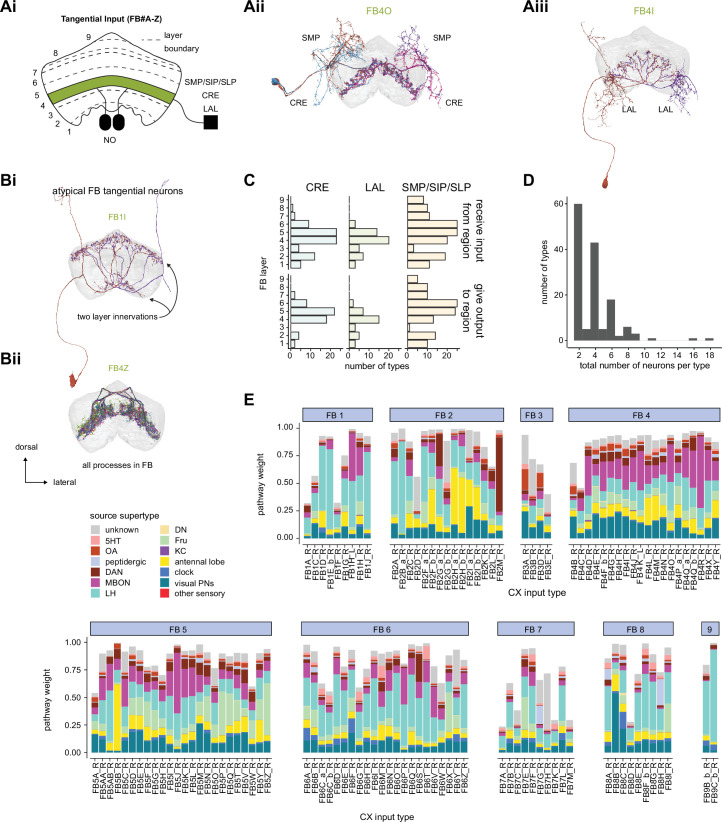
Figure 40. Fan-shaped body (FB) tangential overview.
(A) Overview of FB tangential neurons. (Ai) FB tangential neurons output in single or multiple layers of the FB (e.g., in layer 4, shown in green) and may have mixed arbors in the noduli (NO), superior protocerebrum SMP/SIP/SLP, lateral accessory lobe (LAL), crepine (CRE), or other brain regions outside of the central complex. (Aii) A morphological rendering of FB4O neurons, which receive input in the SMP and CRE and output to layer 4. (Aiii) A morphological rendering of FB4I neurons, which receive input in the LAL and output to layer 4. (B) While most FB tangential neurons arborize in one FB layer and receive input external to the central complex (CX), there are exceptions. Some FB tangential neurons, for example, arborize in multiple FB layers. (Bi) shows a morphological rendering of one such type, the FB1I neurons. In contrast, some FB tangential neurons arborize exclusively within the FB. (Bii) shows a morphological rendering of one such type, the FB4Z neurons. (C) The number of FB tangential types that receive input from (top) or give output to (bottom) the CRE, SMP/SIP/SLP, or LAL. The FB layer refers to the layer where a given type has the most expansive processes. For this analysis, only the right neurons of the type are considered, and each right neuron of that type must have, on average, at least three synapses in the given region. (D) The number of neurons per FB tangential type. With a few rare exceptions, both the right and left FB contribute an equal number of neurons to each type. (E) Input pathway classifications for the FB tangential input neurons. Types are counted as inputs if they have at least 20 synapses of a given polarity outside of the CX and are the postsynaptic partner in at least one significant type to type connection outside of the CX. See Appendix 1—figure 3 for an explanation of pathway weight.


