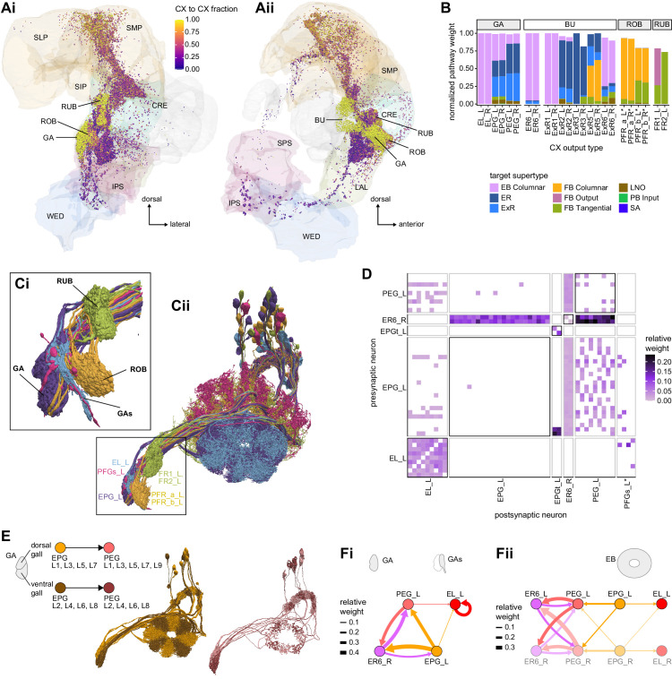
Figure 56. Central complex (CX) to CX connections in the gall (GA), bulb (BU), round body (ROB) and rubus (RUB).
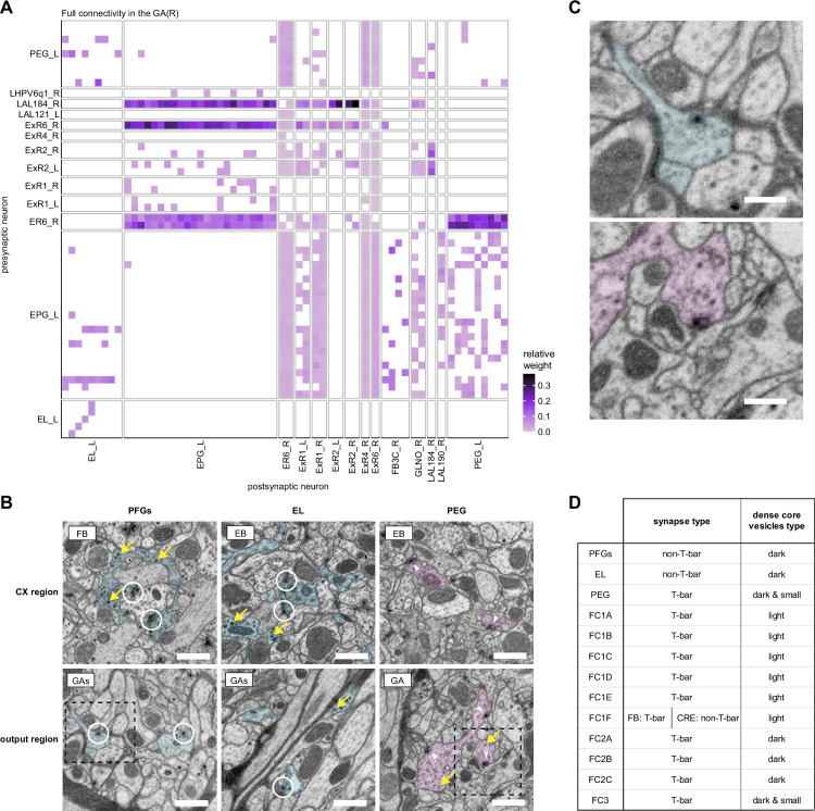
(A) Downstream synapses of potential CX output neurons, colored by the fraction of their target pathways that contribute to pathways coming back to the CX (see Materials and methods). The GA, BU, ROB, and RUB contribute most of their outputs to the CX and the lateral accessory lobe (LAL) almost none, while the upper neuropiles are mixed. (Ai) Frontal view; (Aii) side view. (B) Pathway weights of all pathways that start in the GA, BU, ROB, and RUB and end on another CX neuron. The weights are normalized for each type of origin. If the normalized pathway weight is 1, it corresponds to a neuron for which all output pathways come back to the CX. Connections are separated by the supertypes that these recurrent pathways reach. EB neurons mostly reach other EB neurons, whereas FB neurons mostly reach other FB neurons. (C) Morphological renderings of seven selected neuron types that innervate the GA, gall surround (GAs), ROB, and RUB. (Ci) Closeup of the four structures. (Cii) Illustration of the full morphology of the seven neuron types, showing the left population only. (D) Connectivity matrix of neurons that arborize in the right GA and GAs region. All connections outside of the CX regions (ellipsoid body [EB], protocerebral bridge [PB], fan-shaped body [FB], noduli [NO]) were considered because the GA region of interest does not capture the GAs. PFGs neurons were included in the analysis, but did not make any significant connections. (E) Illustration of selective connectivity between EPG neurons to PEG neurons from odd and even wedges of the EB in the dorsal and ventral GA, respectively. Left: schematic. Middle: rendering of EPG cells targeting the right GA with those from odd wedges colored in orange and those from even wedges colored in brown. Right: rendering of PEG neurons shown analogously as EPG cells (maroon: even-numbered PB glomeruli; pink: odd-numbered PB glomeruli). (F) Connectivity graphs on the level of neuron types showing any connections with at least 0.05 relative weight. (Fi) Connectivity in the GA and GAs. (Fii) Connectivity in the EB between the same neuron types as in (Fi), including the neurons from the other hemisphere.


