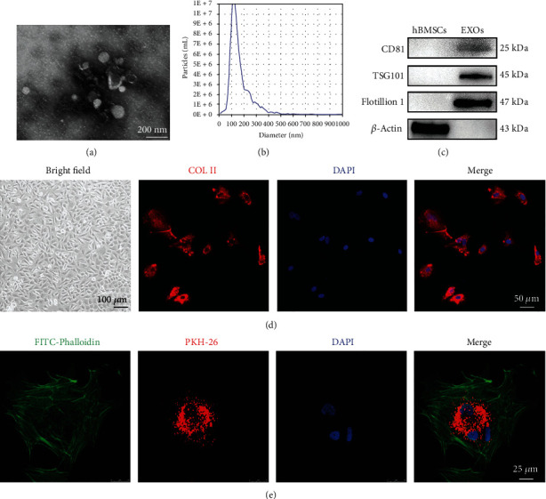
Figure 1.

Characterization of human bone marrow stromal cell- (hBMSC-) derived exosomes (EXOs). (a) Representative transmission electron microscope (TEM) images of hBMSC-EXOs (scale bar = 200 nm). (b) Nanoparticle tracking analysis for hBMSC-EXOs. (c) Western blots showing the exosome markers including CD81, TSG101, and Flotillin 1. β-Actin was used as a negative control. (d) Identification of rabbit chondrocytes. Cell morphology was observed at bright field with a microscope (scale bar = 100 μm). Collagen II (COL II; red) was analyzed by immunofluorescence. DAPI (blue) was used to designate the nuclei. Scale bar = 50 μm. (e) Internalization of hBMSC-EXOs in primary rabbit chondrocytes. hBMSC-EXOs were labeled by PKH26 (red) and incubated with primary chondrocytes. FITC-Phalloidin (green) and DAPI (blue) were used to label the cytoskeleton and the nucleus, respectively. Scale bar = 25 μm.
