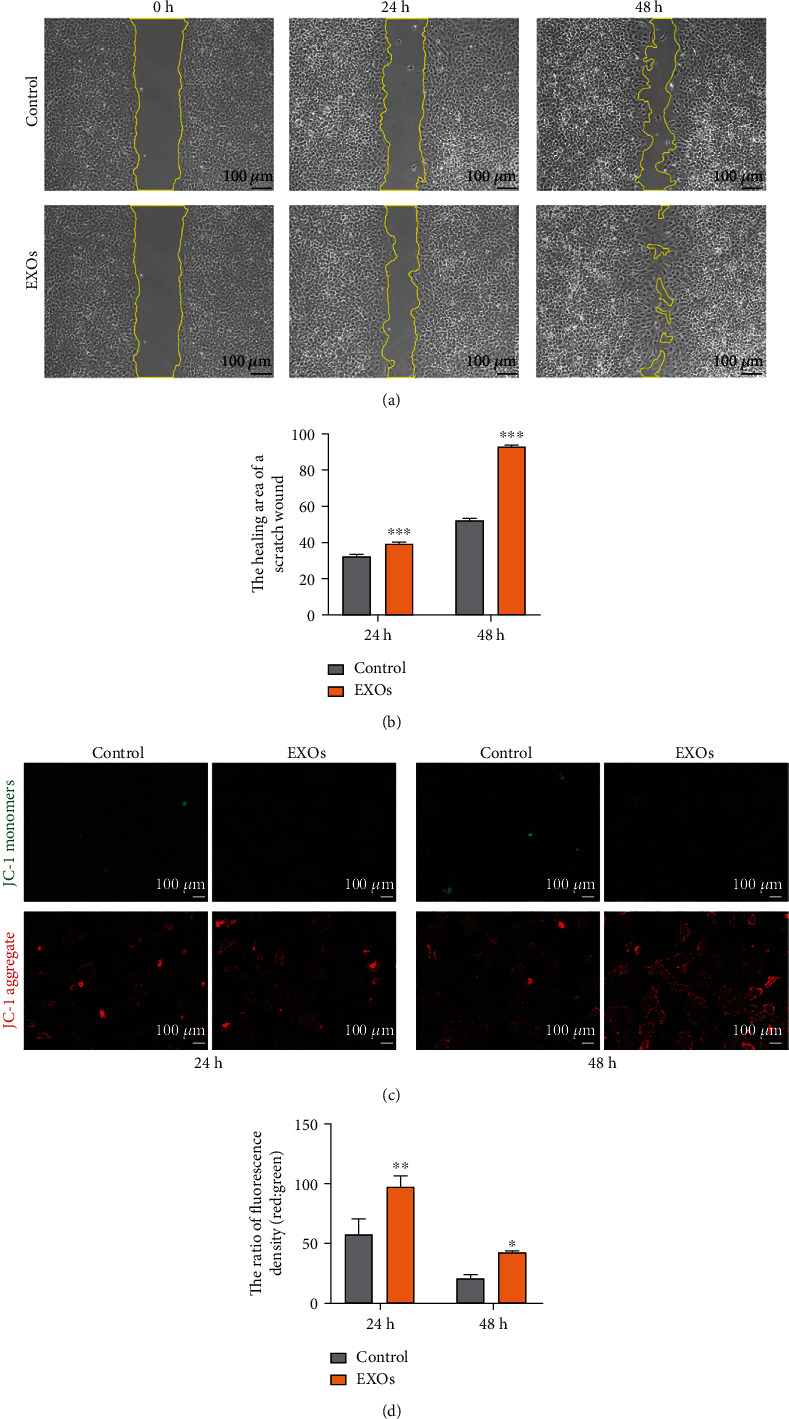
Figure 3.

hBMSC-EXOs promote cell migration and mitochondrial function in chondrocytes. Rabbit primary chondrocytes were treated with hBMSC-EXOs at 1.0 × 10⁹/ml for 24 h or 48 h. Cell migration and mitochondrial membrane potential were assayed. (a) hBMSC-EXOs stimulate cell migration. Wound healing assay was used to evaluate cell migration. Scale bar = 100 μm. (b) Quantitative data for wound healing assay as shown in (a). (c) hBMSC-EXOs increase the mitochondrial membrane potential. Representative fluorescence images for JC-1 staining were shown. Scale bar = 100 μm. (d) Quantitative analysis for JC-1 staining as shown in (c). n = 3. Values are presented as mean ± SEM. ∗P < 0.05, ∗∗P < 0.01, and ∗∗∗P < 0.001, versus the control group, one-way ANOVA.
