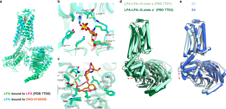
Fig. 7. Structural comparison of LPA1 bound to LPA and ONO-0740556.
a Superimposition of the LPA- and ONO-0740556-bound LPA1 structures, colored green (PDB 7TD0) and cyan, respectively. b, c Superimposition of the binding pocket for LPA and ONO-0740556 in polar regions on the extracellular side (b), and in the hydrophobic pocket (c). d Superimposition of LPA-LPA1-Gi states a (PDB 7TD1) and a’ (PDB 7TD2) aligned at the receptor. e Superimposition of S3 and S4 aligned at the receptor.

