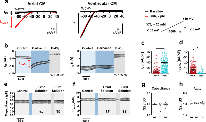
Fig. 5. Inward rectifier acquisition from native atrial and ventricular cardiomyocytes (CM) using automated patch-clamp.
a Representative traces of membrane current (IM) showing basal inward rectifier current (IK1) in atrial and ventricular (vent.) with superimposed acetylcholine-activated inward rectifier (IK,ACh) current following carbachol (CCh) application during a depolarizing ramp voltage protocol. b Time course of a single plate with atrial (n = 10) and ventricular (n = 11) CMs inward current at −90 mV during a typical experiment. Red arrow indicates peak IK,ACh. c Peak inward IK1 density measured at −90 mV. d Peak inward IK,ACh density measured at −90 mV. e Time course of membrane capacitance from a single plate (b, [n = 10]) over various external solution changes. f Time course of series resistance (Rseries) from a single plate (b, [n = 10]) over various external solution changes. g Ratio of mean capacitance changes per plate between solution change 2 (S2) and solution change 3 (S3) over three separate experimental days (D1, D2, D3). h Ratio of Rseries between S2 and S3 over three separate experimental days. Data are mean ± SEM. *P < 0.05 vs. ventricular. n = number of atrial (151) and ventricular (143) CMs from three animals (c, d).

