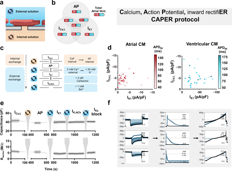
Fig. 6. Overview of the multi-current Calcium, Action Potential and inward rectifiER (CAPER) protocol in native atrial and ventricular cardiomyoctes (CM) using automated patch-clamp.
a Schematic of the cardiomyocyte-aperture interface with corresponding external (blue) and internal (orange) solutions. The substitution of which allows for multi-current acquisition from a single cell. b Shares of total CM numbers that showed successful measurements of any of the three parameters of interest (L-type calcium current (ICa,L), action potential (AP) duration at 90% repolarization (APD90) and basal inward rectifier current (IK1). The central total indicates the successful cohort of atrial and ventricular (Vent.) CMs in which all three parameters were successfully acquired. c A detailed overview of the CAPER protocol including sequential internal and/or external solution exchange during a single experimental run. The acetylcholine-activated inward rectifier current (IK,ACh) is an optional addition to the protocol prior to the inward rectifier block with BaCl2. d Three-dimensional visualization of ICa,L, APD90, and IK1 relationships in atrial (left) and ventricular (right) CMs. APD90 is expressed as a shadow spectrum where darker colors indicate longer AP duration. e Time course of membrane capacitance (upper) and series resistance (Rseries; lower) from a single plate (n = 5 CMs) over a full CAPER run. f Direct acquisition software screenshots from a single plate (single animal) showing three complete electrophysiological measurements from single cells. Y-axis indicates the membrane current expressed as picoamperes (p) or nanoamperes (n) in the screenshots of ICa,L (left) and IK1 (right), or membrane voltage expressed as millivolts (m; center).

