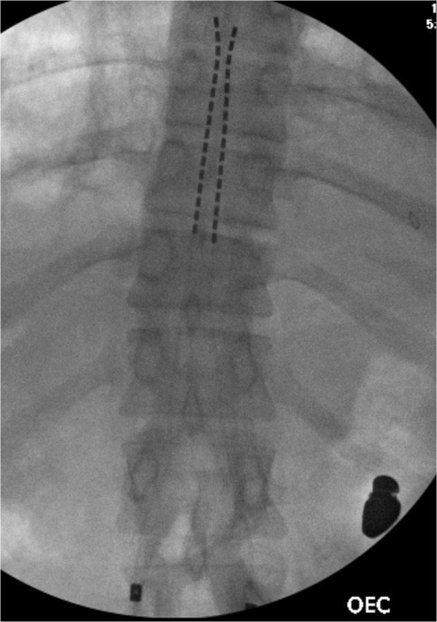
Fig. 5. Intra-procedural AP fluoroscopic image of the lower thoracic spine.

This demonstrates placement of the spinal cord stimulation leads starting at the superior aspect of the T11 vertebral body, and terminating at the inferior aspect of the T8 vertebral body. A bullet fragment is also visualized.
