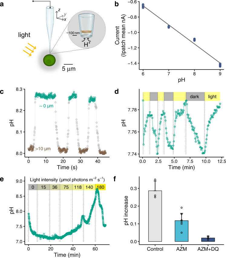
Fig. 1. Phycosphere pH of marine green algae Chlamydomonas concordia RCC1 (~5 µm diameter).
a A schematic showing the operation of a pH sensing nano-probe for in situ measurement of pH in the phycosphere of a single cell C. concordia by the scanning ion conductance microscopy. b The relationship between the measured ion current of the pH nano-probe and seawater pH (n = 12, r2 = 0.99, p < 0.0001). c A significant pH rising when closing to an illuminated cell. The difference in the pH at the cell surface (8.27 ± 0.01, ~0 µm away from the cell) and that at >10 µm measuring point (8.01 ± 0.01) was significant (p < 0.0001). The flagella-mediated motility of C. concordia reduced the spatial resolution of local pH profile. Bulk seawater pH = 8.00. d A representative pattern for the pH change in the phycosphere under consecutive light/dark cycles. Bulk seawater pH = 7.74 and HCO3− = 0.4 mM, the low concentration of HCO3− was used to facilitate the measurement of light/dark effect. e The pH in the phycosphere increased with increasing light intensity, and at the highest light intensity the pH decreased due to possible photosynthesis inhibition. Bulk seawater pH = 7.92, HCO3− = 0.4 mM. f The increase in the phycosphere pH in illuminated cells was significantly inhibited by 100 μM acetazolamide AZM (inhibitor of external carbonic anhydrase, n = 8, p = 0.000), and the pH increase was completely suppressed (n = 3, p = 0.074 in comparison with 0.00) following further addition of 8 μM diquat dibromide DQ (inhibitor of photosystem I). Bulk seawater pH = 7.93 and HCO3− = 0.4 mM.

