Fig. 4.
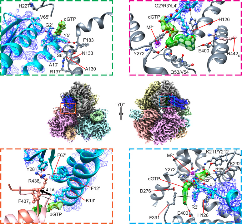
Interactions between Dgt, Gp1.2, and dGTP. Middle, overview of the Dgt–Gp1.2–dGTP structure. Dgt and Gp1.2 are colored by monomer, while the dGTPs are colored green. Top Left, close-up of hydrogen-bond interactions between Gp1.2 and Dgt monomer A. Bottom Left, close-up of cation–π interactions between Gp1.2 and Dgt monomer E. The apo-Dgt (PDB ID: 4XDS, colored salmon) is aligned by the monomer A active-site histidine-aspartate (HD) motif. A portion of monomer E is displaced by >4 Å (as measured by the R436E C-α) to accommodate Gp1.2 helix α1. Top Right, close-up of interactions between Gp1.2, Dgt monomer A, and dGTP. Bottom Right, point of view rotated ∼90° from the image in the Top Right. The Middle images were generated in ChimeraX, while the other images were generated in Chimera.

