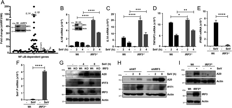
Fig. 3.
IRF3 inhibits virus-induced inflammatory gene induction in human cells. (A) Graphical presentation of the microarray analyses of the NF-κB–dependent genes in Wt and shIRF3 (immunoblot of IRF3 expression; inset in A) HT1080 cells after SeV infection (2 h postinfection), are as described in Methods; the genes are listed in SI Appendix, Tables S1 and S2. The microarray results are from duplicate samples for each condition. (B–F) Wt or IRF3−/− (immunoblot of IRF3 expression; inset in B) HT1080 cells were infected with SeV (B–F), and the mRNAs of inflammatory target genes (B–E) or viral mRNA (F) were analyzed by qRT-PCR. (G) Wt and IRF3−/− (knockout [KO]) HT1080 cells were infected with SeV for the indicated time, and the cell lysates were analyzed for TNFAIP3/A20, IFIT1, and IRF3 by immunoblot. (H) The IRF3 shIRF3 HT1080 cells were infected with SeV for the indicated time, and the cell lysates were analyzed for TNFAIP3/A20 and IFIT1 by immunoblot. (I) Wt and IRF3hi U4C cells were infected with SeV and analyzed for TNFAIP3/A20 (Upper panel) and IRF3 (Lower panel) by immunoblot. NT, nontargeting. The results are representative of three experiments; the data represent mean ± SEM. SeV P, SeV P gene, *P < 0.05, **P < 0.005, ***P < 0.0005, ****P < 0.0001.

