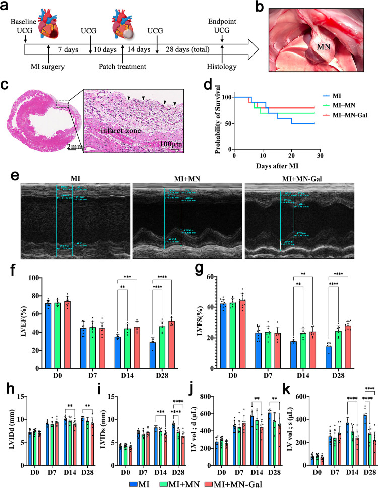
Figure 2.
MN-Gal patch preserved the cardiac structure and function following MI. (a) Schematic showing the overall animal study design used to test the therapeutic benefits of MN-Gal patch in a rat model of MI. (b) Placement of an MN patch on the rat heart. (c) H&E staining indicates the MN pinholes (black arrow) on the infarcted and peri-infarct zone of the infarcted heart (scale bars: 2 mm, left; 100 μm, right). (d) Survival curve of MN-Gal patch-treated MI rats compared with that of blank MN patch-treated MI rats and only MI rats. (e) Representative M-mode echocardiographic image showing the LV wall motion of the hearts 28 days after MI. Diastolic and systolic cycles were analyzed for each image, and three images for each time point were analyzed per rat. (f–k) LVEF, LVFS, LV internal dimension at end-diastole (LVID; d) and end-systole (LVID; s), and LV volume at end-diastole (LV Vol; d) and end-systole (LV Vol; s) were measured by echocardiography before MI surgery (baseline) and 7, 14, and 28 days (endpoint) after MI. n ≥ 5 animals per group. All data were presented as means ± SD. Comparisons between three groups were performed using two-way ANOVA, followed by Tukey’s multiple comparison test. **P < 0.01; ****P < 0.0001 between each group and every other group.

