Figure 1.
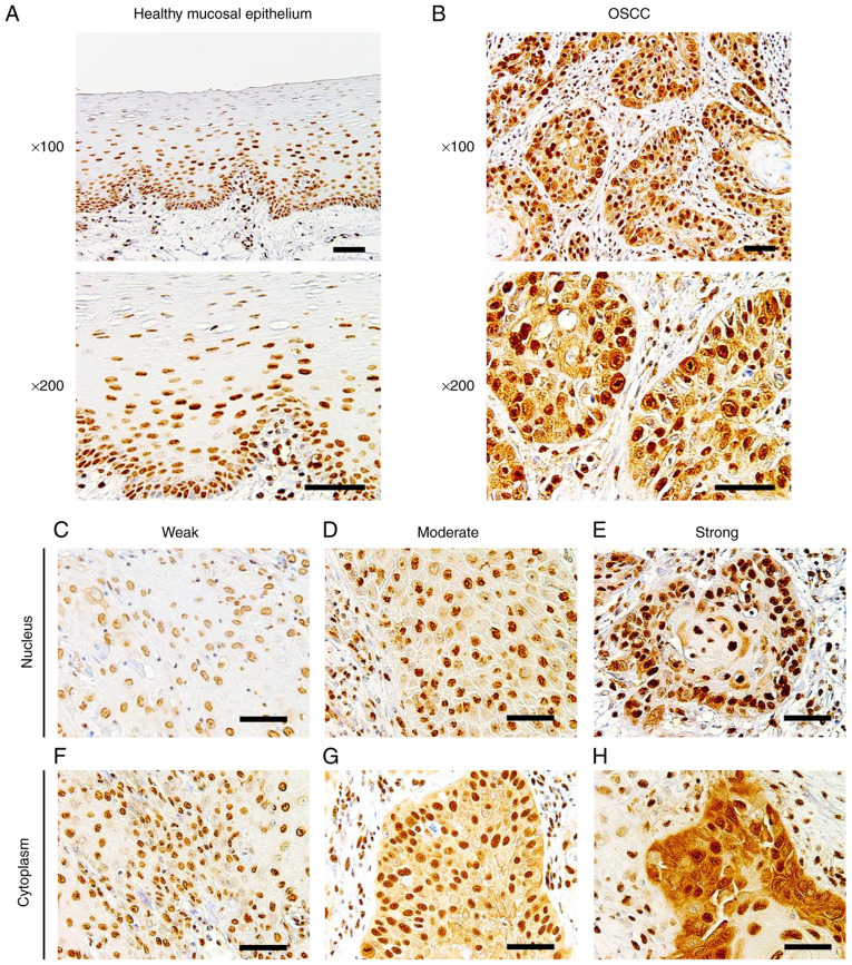
Immunohistochemistry of Sam68. (A) Representative images of healthy mucosal epithelium adjacent to OSCC, in which Sam68 expression was predominantly detected in the nucleus and negatively observed in the cytoplasm (magnification, ×100 and ×200; scale bar, 50 µm). (B) Representative image of OSCC, in which Sam68 expression was detected both in the nucleus and cytoplasm (magnification, ×100 and ×200; scale bar, 50 µm). (C-E) Nuclear expression of Sam68 with (C) weak, (D) moderate, and (E) strong staining intensity in OSCC cells (magnification, ×200; scale bar, 50 µm). (F-H) Cytoplasmic expression of Sam68 with (C) weak, (D) moderate, and (E) strong staining intensity in OSCC tissue (magnification, ×200; scale bar, 50 µm). Sam68, Src-associated in mitosis 68 kDa; OSCC, oral squamous cell carcinoma.

