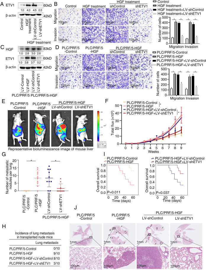
Fig. 6.
ETV1 is vital for HGF-mediated HCC invasion and metastasis. (A) The protein levels of ETV1 in the PLC/PRF/5 cells transfected with LV-sh Control or LV-shETV1 upon HGF treatment. (B) Transwell assays displayed the migratory and invasive capacity of the indicated cells upon HGF treatment. (C) The levels of HGF and ETV1 in the PLC/PRF/5-HGF cells transfected with LV-shControl or LV-shETV1. (D) Transwell assays displayed the migratory and invasive capacity of the indicated cells. (E-J) In vivo metastatic assay. (E) The representative BLI images in the liver were shown 9 weeks after implantation with indicated cells. (F) The bioluminescent signals were used to show the growth rate of liver tumors. (G) The number of metastatic lesions in the lung tissues. (H) The occurrence of lung metastasis. (I) The OS of different groups of nude mice. (J) Representative images of H&E staining of lung samples (indicated by arrowheads) from each group. * Represented p < 0.05. All data were displayed as Mean ± SD

