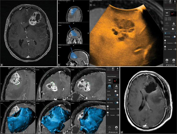
Figure 2:
Patient 1. Preoperative axial MRI with contrast showing the extent of tumor involvement in the left frontal lobe (a). Intraoperative Images showing the integration of real-time ultrasound images with the Brainlab navigation system (b). Using real-time ultrasound to safely and efficiently determine the extent of resection of the tumor bed and effectively accounting for possible brain shift towards the end of surgery (c). Postoperative axial MRI with contrast showing complete tumor resection (d).

