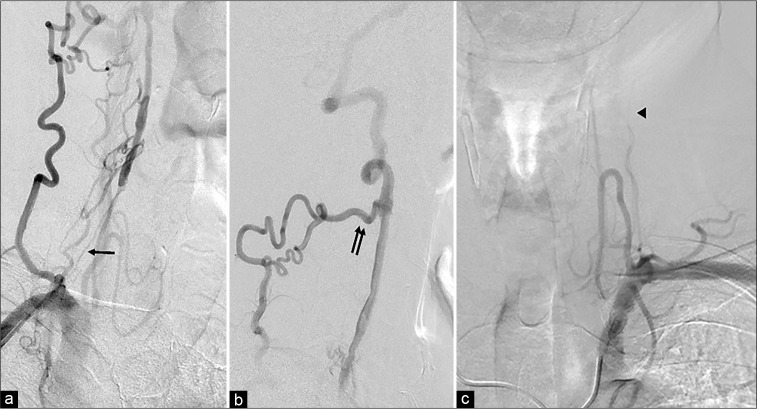
Figure 2:
(a) The right anterior oblique view of the vertebral artery (VA) angiogram shows pseudo-occlusion of the right VA orifice with faint anterograde flow (arrow). (b) Lateral view of the right VA lateral angiogram reveals that the right VA is reconstructed with a collateral pathway through the deep cervical arteries (double arrows). (c) The left subclavian angiogram reveals left VA occlusion at the C2 level (arrowhead).

