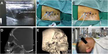
Figure 2.

The process flows of PRF.
Note: (A) The transverse ultrasound image of T4. (B) The upper and target DRGs (T3 and T4 DRGs) were stimulated for 15 minutes. (C) The target and lower DRGs (T4 and T5 DRGs) were stimulated for 15 minutes. The T4 DRG was the primarily involved DRG and was stimulated for 30 minutes. (D) A lateral 3D C-arm reconstruction image showing the position of the needle in foramen rotundum. (E) A lateral C-arm image showing the position of the needle in foramen ovale. (F) PRF was ongoing in the patient. DRG: Dorsal root ganglion; PRF: pulsed radiofrequency.
