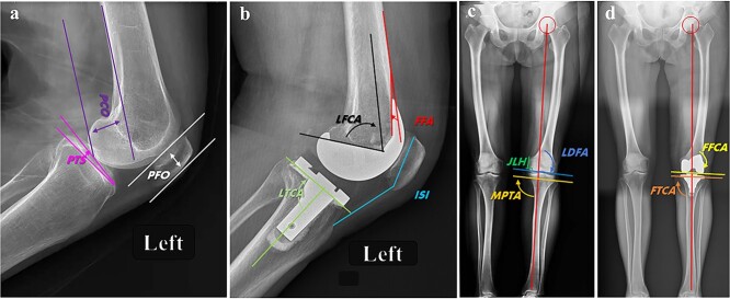
Figure 2.
Illustrative index of radiographical measurement before and after TKA. a, b are the preoperative and postoperative lateral radiographs of the knee X-ray at 30° of flexion for evaluation of components position. PCO is the distance between the posterior femoral condyle and the tangent line of the posterior femoral cortex; PTS is the angle between the tibial plateau and the horizontal plane; PFO is the distance between the trochlear groove of the femur and the straight line of the anterior patellofemoral cortex; LFCA is the angle between the longitudinal axis of the femur and the prosthesis in sagittal plane; LTCA is the angle between the longitudinal axis of the tabia and the prosthesis in sagittal plane; FFA is the angle between the tangent line of the anterior femoral cortex and the anterior condyle of the prosthesis; ISI is the ratio of patellar height and patellar tendon length; JLH was determined by the distance from the prominence of medial condyle to the distal medial condyle of femur. c. the preoperative standing full-length radiographs of left lower extremity showed a moderate genu varus deformity with degenerated LDFA (91.5°), and MPTA (90.2°). d. postoperative standing full-length radiographs showed the lower limb alignment was ideally corrected as 180.0° in mechanical axis, with a FFCA 90.0°, FTCA 87.0°. TKA total knee arthroplasty, PCO posterior condylar offset, PTS posterior tibial slope, PFO patellofemoral offset, LFCA lateral femoral component angle, LTCA lateral tibial component angle, FFA femoral flexion angle, ISI Insall-Salvalti index, JLH joint line height, LDFA lateral distal femoral angle, MPTA medial proximal tibial angle, FFCA frontal femoral component angle, FTCA frontal tibial component angle

