Figure 4.
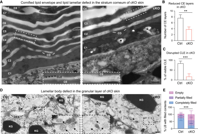
Ultrastructural analysis reveals KLF5 is required for cornified envelope formation and lipid secretory functions. (A) TEM at 25,000× magnification shows fewer and largely disorganized cornified envelope (CE) layers from cKO epidermis (right, C1–C4 cornified layers) compared with Ctrl (left, C1–C8 cornified layers). Dashed line insets show magnified regions from Ctrl and cKO samples and demonstrate in the Ctrl sample well-organized electron-dense CE, attached by a lucent band of cornified lipid envelope (CLE), and further sandwiched by translucent intercellular lipid lamellae (LL). CLE and LL are significantly disrupted in cKO. Asterisks denote abnormal lipid aggregates found in the cKO stratum corneum, reminiscent of case reports from skin congenital disorder patients with barrier defects. (CD) Corneodesmosome, (Der) dermis. Scale bar, 500 nm. (B,C) CE (B) and CLE (C) are quantified. (D) TEM at 25,000× magnification shows the granular layer harboring keratohyalin granules (KG), keratin filaments (KF), and desmosomes (DES). Lamellar bodies (black arrows) show lipid stacks in the Ctrl group but appear largely vacant from cKO. Scale bar, 100 nm. (E) Lamellar bodies are quantified. Unpaired t-tests were performed for B, C, and E. Data are mean ± SEM. (**) P < 0.01, (***) P < 0.001.

