Figure 5.
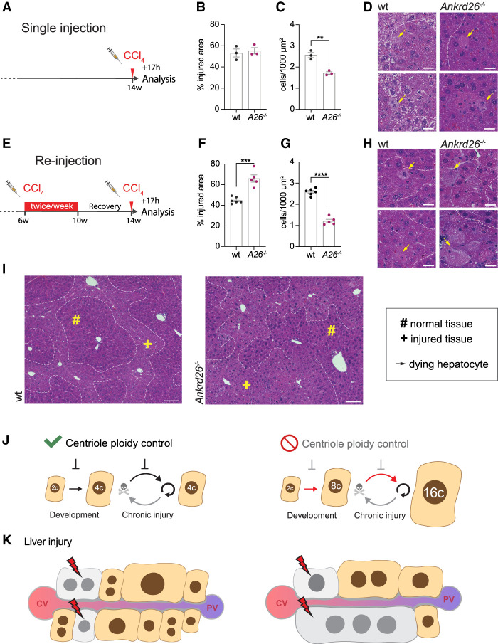
Massive ploidy increases sensitize the liver to injury. (A) Treatment scheme for single injection: Fourteen-week-old male mice were injected with a single dose of 5 mL/kg CCl4 and analyzed after 17 h. (B,C) The injured area (B) and cell density in this region (C) were quantified in H&E-stained wt and Ankrd26−/− (A26−/−) liver sections. N = 3 mice per genotype. (D) Representative close-ups of H&E-stained liver sections show healthy and affected liver tissue separated by dashed lines. Yellow arrows mark dying hepatocytes. Scale bar, 10 µm. (E) Treatment scheme reinjection: Six-week-old male mice were injected biweekly with 5 mL/kg CCl4 for 4 wk. Following 28 d of recovery, the mice were reinjected with a single dose of 5 mL/kg CCl4 and the livers were harvested after 17 h. (F,G) Quantification of the affected area (F) and the cell density within the injured region (G) performed on H&E-stained liver sections also shown in H and I. For wt, N = 6. For Ankrd26−/−, N = 5. (H) Close-ups of wt and Ankrd26−/− livers. Dashed lines separate normal and injured tissue, and yellow arrows indicate dying hepatocytes. Scale bar, 10 µm. (I) Tissue overview of wt and Ankrd26−/− livers treated as shown in E. Scale bar, 100 µm. (#) Intact tissue, (+) affected tissue. (J,K) Model summarizing how centriole-directed ploidy control maintains a low degree of polyploidy during development and chronic injury cycles. The absence of this pathway results in hyperpolyploidy over multiple cycles of compensatory proliferation. (K) Additional injury targeting the same number of cells impacts the hyperpolyploid liver to a greater extent. (CV) Central vein, (PV) portal vein. All data are shown as mean ± S.E.M and statistical significance was determined using an unpaired two-tailed Student's t-test. Only significant results are indicated. (**) P < 0.01, (***) P < 0.001, (****) P < 0.0001.

