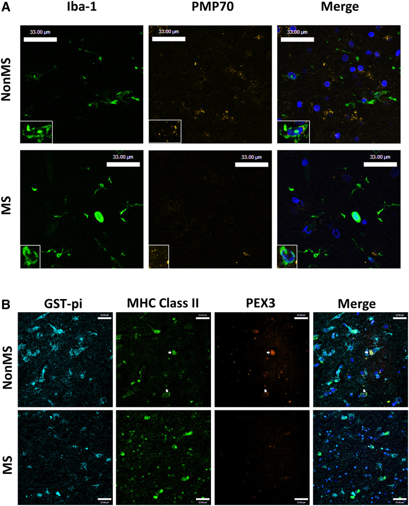
Figure 2.
PMP70 and PEX3 immunoreactivity is suppressed in subcortical white matter of patients with MS. A, Representative immunofluorescence images of white matter from MS patients and non-MS controls labeling Iba-1 (green), PMP70 (yellow), and DAPI (blue) with high-magnification inset. B, Immunofluorescence image of subcortical white matter from a MS patient compared with a non-MS patient labeling GST-π (cyan), MHC II (green), PEX3 (orange), and DAPI (blue). Arrows point to MHC Class II positive cells with PEX3 immunostaining. Scale bars, 33 µm.

