
FIGURE 1.
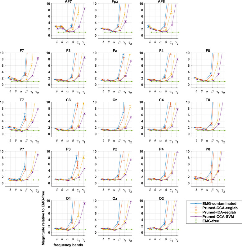
Montage showing extent of EMG contamination of EEG relative to EMG‐free recording with different pruning methods. Divergence from EMG‐free is prominent in gamma bands and increased with increasing distance from the central scalp at Cz. Delta and theta bands show small increases, while alpha bands are reduced during paralysis (see also Figure 3).