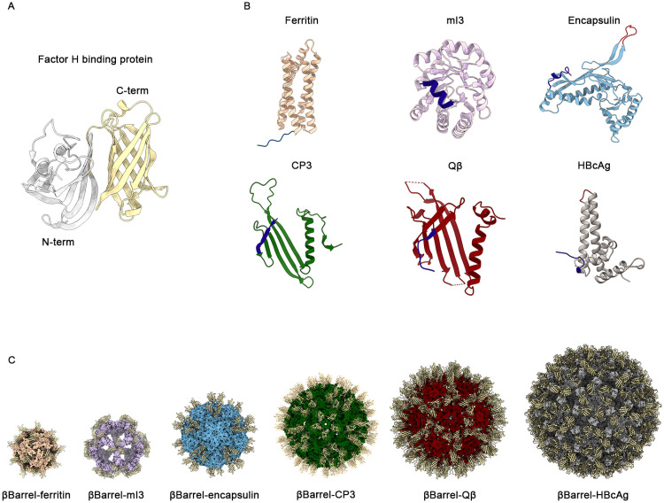
Fig 1. Structural analysis and in silico design of chimeric NPs.
(A) Cartoon representation of 3D structure of fHbp antigen (pdb code 3KVD). In grey it is reported the N-terminal domain (residues 1–118) while in yellow it is shown the C-terminal βbarrel domain used in this work (residues 119–249). (B) Cartoon representation of the monomeric structure of each tested NPs. Engineerable sites explored for the genetic fusion of the antigen are highlighted: the N terminus in dark blue and the exposed loops in red. (C) Cartoon of predicted 3D models of each chimera obtained with Rosetta homology modelling. The βbarrell exposed was represented in yellow. Images were obtained with ChimeraX (panel A, C) and Pymol (panel B).

