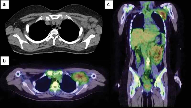
Figure 1.

Chest computed tomography (CT) and gallium-67 single-photon emission-CT (SPECT) scintigraphy. (a) Chest CT on admission shows multiple enlarged lymph nodes in the left axilla. (b–c) Gallium-67 SPECT scintigraphy shows strong uptake in the axillary lymph nodes. No lymph node swelling or uptake is detected in other parts of the body.
