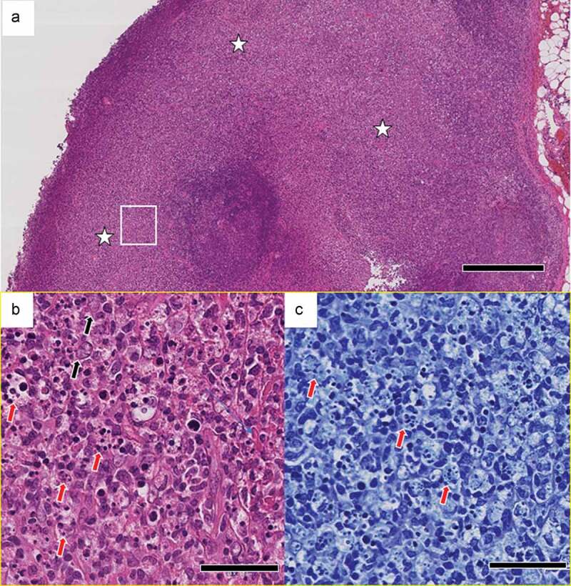
Figure 2.

Pathological findings in the left axillary lymph node. (a) Localized, focal lesions (white stars) that obscure the lymph node structure. [hematoxylin & eosin (H&E)]. Scale bar: 500 μm. (b) Numerous nuclear debris (red arrows) and some enlarged lymphocytes (black arrows). High-magnification view of the area in the white rectangle in (a). Scale bar: 50 μm. (c) Granulocytes are not evident. Nuclear debris (red arrows) that appear to have been phagocytosed by histiocytes. High-magnification view of the area in the white rectangle in (a). [Giemsa stain.] Scale bar: 50 μm.
