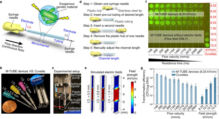
Fig 1. M-TUBE is a fabrication-free, microfluidics tubing-based bacterial electroporation device that is simple to assemble and exhibits higher electroporation efficiency than cuvettes.
(a) Schematic of the M-TUBE device. The device is composed of 2 syringe needles and 1 piece of plastic tubing of predefined length. The 2 syringe needles and plastic tubing serve as the 2 electrodes and microchannel, respectively. When the 2 electrodes are connected to an external power supply (or electrical signal generator), an electric field is established within the microchannel, where bacterial electroporation can take place. (b) M-TUBE devices with 3 ID are all similar in size to a conventional cuvette. (c) Photograph of the experiment setup when using the M-TUBE device. Since the M-TUBE device is made from standard, commercially available syringe needles and plastic tubing, it can be readily attached to syringe pumps for automated sample delivery, removing the need for manually pipetting samples. (d) Detailed breakdown of the protocol for M-TUBE assembly. One device can be completely assembled in 90–120 s. The total cost of parts is currently less than $0.22 and this price could be lowered if parts are bought in bulk. (e) Simulations of the electric field established in M-TUBE devices using COMSOL Multiphysics predict similar field strengths irrespective of ID. (f) Spot-dilution assay to quantify viability on selective plates when E. coli NEB10β cells were flowed through the device with a plasmid encoding ampicillin resistance and GFP (S4 Table) in the presence or absence of an electric field. Transformation was dependent on the electric field. For M-TUBE devices, a voltage of ±2.50 kV (AC field) was applied, which results in an electric field of 8.33 kV/cm. The same batch of cells was used to conduct cuvette-based electroporation as a comparison. (g) Comparison of transformation efficiency (CFUs per μg of DNA) corresponding to the plates in (f). The electroporation efficiency of M-TUBE decreased as the fluid velocity was increased, as expected due to the shorter duration of exposure to the electric field. Regardless of the fluid velocity, the efficiency of M-TUBE was at least 1 order of magnitude higher than that of cuvettes with the same field strength (8.33 kV/cm). Data represent the average (n ≥ 3) and error bars represent 1 standard deviation. The data underlying Fig 1E and 1G can be found in S1 and S2 Data files, respectively. AC, alternating current; CFU, colony-forming unit; GFP, green fluorescent protein; ID, inner diameter; M-TUBE, microfluidic tubing-based bacterial electroporation.

