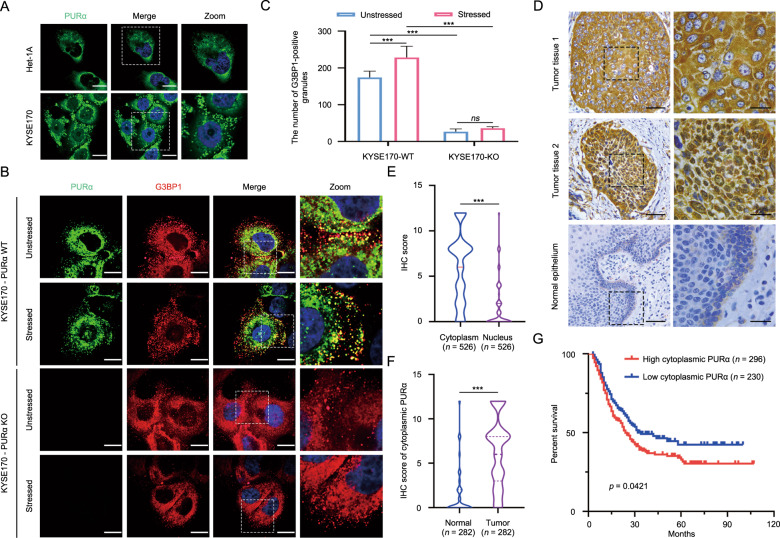
Fig. 1. Cytoplasmic PURα participates in the formation of stress granules and significantly correlates with ESCC progression.
A The localization of endogenous PURα in esophageal epithelium Het-1A and ESCC cancer (KYSE170) cells was visualized by immunofluorescence assay. PURα proteins dispersed in the cytoplasm as granules in KYSE170 cells or accumulated around the nucleus in Het1A cells. Scale bars: 30 μm. B The colocalization between endogenous PURα and the stress granule maker G3BP1 was visualized in wild-type (WT) and PURα-deficient KYSE170 (KO) cells by immunofluorescence staining. Scale bars: 30 μm. C The number of PURα/G3BP1-positive granules under stress conditions or not was calculated separately. ***p < 0.001; ns, not significant. D PURα expression in ESCC tumor tissues (first and second panels) and adjacent nontumor epithelia (third panel) was compared by immunohistochemical staining (IHC). PURα protein mainly located in cytoplasm (first panel) or nucleus (second panel) is shown. The representative region (black frame) at low magnification (40×, left) was amplified at high magnification (100×, right). Scale bars: 50 μm. E Violin plots of the statistical data regarding the IHC score of PURα protein in the cytoplasm and nucleus of tumor tissues (n = 526). ***p < 0.001 by Mann–Whitney test. F Violin plots of the statistical data regarding the IHC scores for cytoplasmic PURα in ESCC (tumor) and adjacent nontumor (normal) tissues (n = 282) were drawn. ***p < 0.001 by Mann–Whitney. G Kaplan–Meier analyses of overall survival. Patients with high cytoplasmic PURα expression (n = 296) had a significantly lower overall survival rate than patients with low cytoplasmic PURα expression (n = 230).

