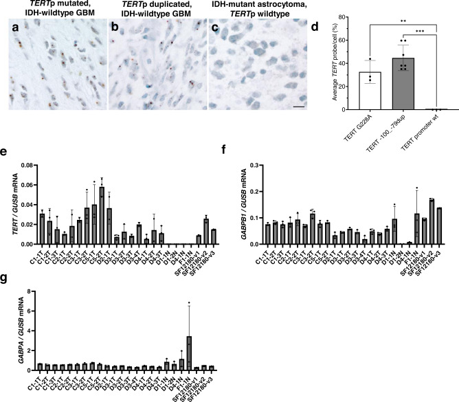
Fig. 5. Similar TERT expression in glioblastomas with TERT promoter duplication or hot-spot mutation.
a–d RNAscope for TERT expression in human glioblastoma. Representative images from RNAscope demonstrating (a) TERT mRNA detected at the single cell level in a positive control glioblastoma, IDH-wildtype with TERT G228A mutation, b TERT mRNA in glioblastoma, IDH-wild type with TERT −100_−79 duplicated (SF12747), (c) TERT mRNA in a negative control IDH-mutant astrocytoma with ATRX mutation and wildtype TERTp. Nuclei stained with hematoxylin. Scale bar denotes 30 µm (a–c). d Quantification of the average TERT probe per total cell number (%) for the 3 tumors illustrated in (a–c). N = 1496 cells examined over 3 independent experiments for TERT G228A, n = 3204 cells examined over 7 independent experiments for TERT −100_−79dup, n = 547 cells examined over 3 independent experiments for TERT promoter wt. (e–g) TERT, GABPB1 and GABPA RT-qPCR results normalized to GUSB and performed on RNA isolated from the multifocal TERTp duplicated tumor (T) (SF12747), adjacent normal (N) (SF12747) and TERTp G228A (SF12180) FFPE punches. Values are mean and SD. Multiple comparisons were performed using 1-way ANOVA, and post hoc analyses were based on Tukey’s test. **p = 0.005, ***p = 0.0001. C/D# indicates the FFPE tissue block and T/N# indicates the punch. Abbreviations: TERTp, TERT promoter. Source data are provided as a Source Data file. N = 3 technical replicates.

