Figure 3.

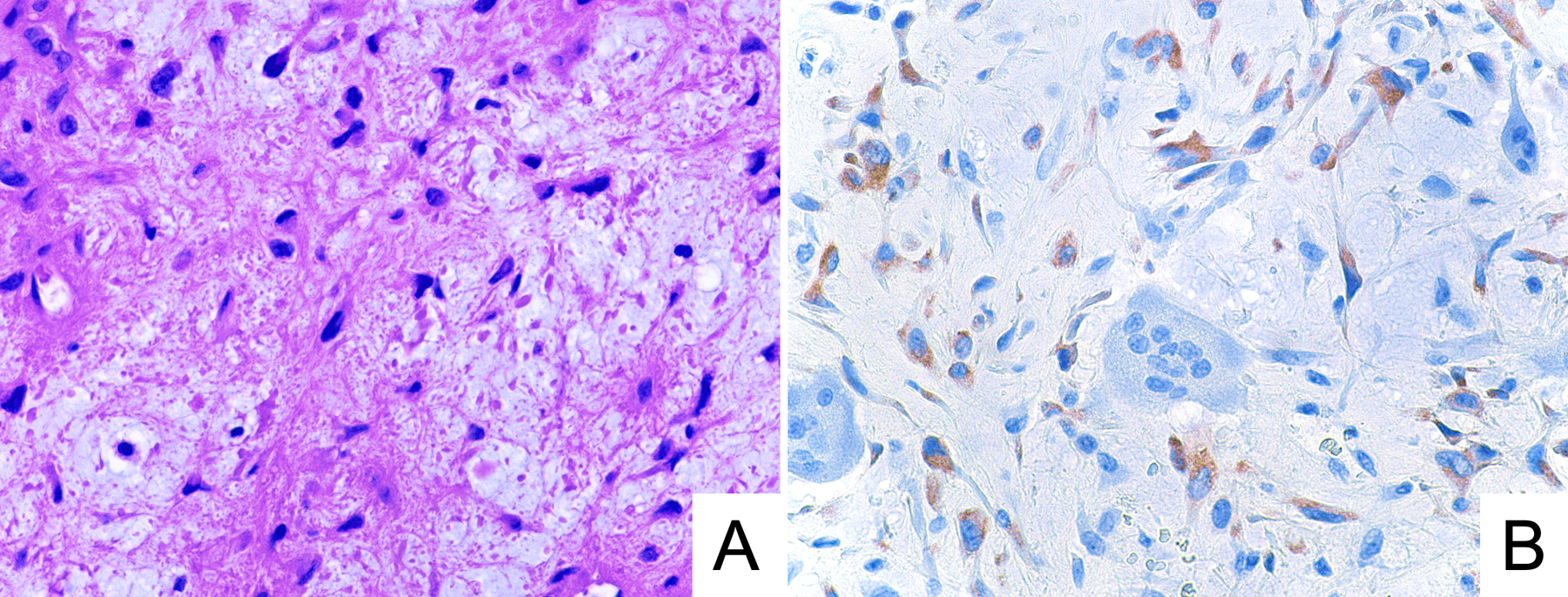
Representative photomicrographs of H&E stain (A) and GRM1 immunohistochemistry (B) in a case of chondromyxoid fibroma exhibiting moderate anti-GRM1 staining intensity. This specimen underwent acid decalcification.

Representative photomicrographs of H&E stain (A) and GRM1 immunohistochemistry (B) in a case of chondromyxoid fibroma exhibiting moderate anti-GRM1 staining intensity. This specimen underwent acid decalcification.