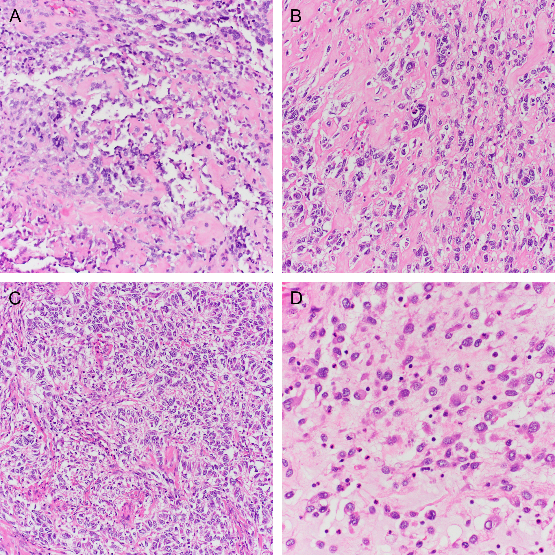
Figure 6.

(A) A scant pretreatment biopsy of a tumor with IRF2BP2-NTRK1 fusion (case 14) with prominent stromal hyalinization. The post-radiation resection specimen showed unusual morphology including (B) atypical epithelioid cells embedded in a hyaline matrix, (C) trabecular architecture, and (D) discohesive rhabdoid cells.
